Figure 2. Retinal morphology of rodless rd1 and rd10 mice, at 24 days of age. Both rodless rd1 (left panel) and rd10 (middle panel) retinas exhibit marked thinning of the photoreceptor layer. Note the normal ONL thickness of the dark-reared
rd10 mouse retina (right panel), in contrast to that of the cyclic light-reared rd10 (middle panel); ONL, outer nuclear layer; IS, inner segments; INL, inner nuclear layer; GCL, ganglion cell layer; Reprinted
from Vision Research, vol. 47(5), Chang B, et al., Two mouse retinal degenerations caused by missense mutations in the beta-subunit of rod cGMP
phosphodiesterase gene, 624-33, 2007, with permission from Elsevier.
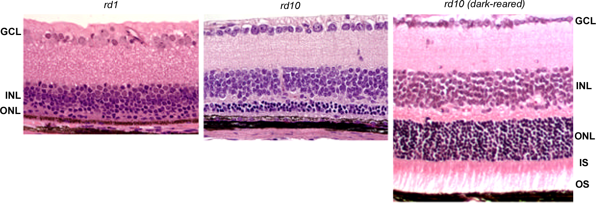
 Figure 2 of
Han, Mol Vis 2013; 19:2579-2589.
Figure 2 of
Han, Mol Vis 2013; 19:2579-2589.