Figure 1. Schematic representation of the mouse PDE6B gene and protein, and the localization of spontaneous mutations in animal models.
The rd1 mouse contains a murine leukemia provirus insertion in intron 1 and a point mutation, which introduces a stop codon in exon
7. The rd10 mouse carries a missense mutation (R560C) in exon 13. Two canine models, the rcd1 Irish setter and the Sloughi dog, contain a nonsense amber mutation at codon 807 (W807ter) and an 8 bp insertion after codon
816, respectively. The PDE6B protein contains two high-affinity non-catalytic cGMP binding sites (GAF domains) and a catalytic
domain in which the majority of human mutations are located. Reprinted from Vision Research, vol. 49(22), Baehr W. and Frederick J.M., Naturally occurring animal models with outer retina phenotypes, 2636–2652, 2009,
with permission from Elsevier.
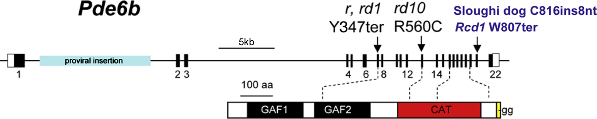
 Figure 1 of
Han, Mol Vis 2013; 19:2579-2589.
Figure 1 of
Han, Mol Vis 2013; 19:2579-2589.