Figure 8. Contractile features of BALB/c mouse aqueous drainage tract. A-C: Reticular pattern of MHC and F-actin labeling was seen in the ciliary body (CB) of BALB/c mice (n=6), which was otherwise
obscured by pigment in C57BL/6 mice. D: MHC was mainly localized in trabecular meshwork (TM; closed arrows) adjacent to Schlemm’s canal (SC; asterisk*). E, I, M: the pattern of α-SMA, caldesmon, and calponin labeling in BALB/c mice was similar to that of C57BL/6 mice, confirming that
pigment did not obscure interpretation of the CM in C57BL/6 mice. H, L, P: TM F-actin labeling in the TM adjacent to SC (asterisk*) did not co-localize with a-SMA, caldesmon or calponin (mainly closed
CM). Scale bars, 20 μm.
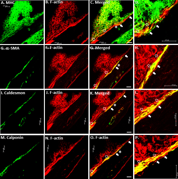
 Figure 8 of
Ko, Mol Vis 2013; 19:2561-2570.
Figure 8 of
Ko, Mol Vis 2013; 19:2561-2570.