Figure 7. Primary immunolocalization of α-SMA, caldesmon, calponin, and MHC in the trabecular meshwork (TM) and ciliary muscle (CM).
A-D: colocalization of α-SMA and caldesmon was primarily in CM (open arrow, yellow). E-H: α-SMA was mainly localized in CM (opened arrow, green), while MHC was mainly localized in the TM (closed arrows) adjacent
to SC (asterisk *). I-L: calponin was mainly localized in CM (opened arrow, green), while MHC was mainly localized in the TM (closed arrows) adjacent
to Schlemm’s canal (SC; asterisk *). Scale bars, 20 μm.
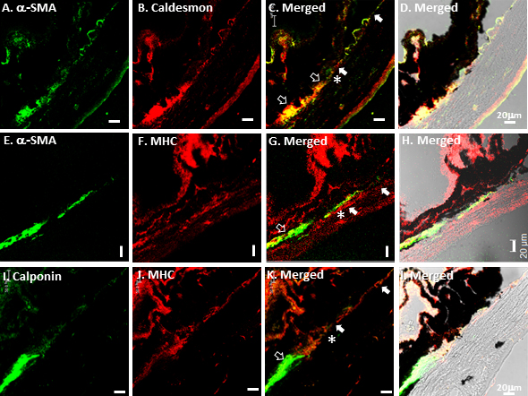
 Figure 7 of
Ko, Mol Vis 2013; 19:2561-2570.
Figure 7 of
Ko, Mol Vis 2013; 19:2561-2570.