Figure 10. Organization of trabecular meshwork (TM) and cilliary muscle (CM) based on foregoing contractile marker localization studies
of F-actin, MHC, α-SMA, caldesmon, and calponin in the mouse aqueous drainage tract. An anterior part of the TM faces the
anterior chamber (anterior TM), while a more posterior part is sandwiched between CM and Schlemm’s canal and does not face
the anterior chamber (posterior TM). The CM extends from the iridocorneal angle of the anterior chamber to the anterior termination
of retina posteriorly.
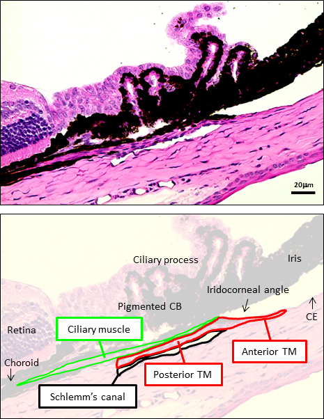
 Figure 10 of
Ko, Mol Vis 2013; 19:2561-2570.
Figure 10 of
Ko, Mol Vis 2013; 19:2561-2570.