Figure 1. Histological features of mouse aqueous drainage tissue based on hematoxylin and eosin (H&E) staining. Enucleated eyes of C57BL/6
mice were embedded in formalin-fixed paraffin embedded sections then H&E stained. Opened arrows indicate ciliary muscle; closed
arrows indicate trabecular meshwork; asterisk (*) indicates Schlemm’s cannal. Scale bars, 20 μm.
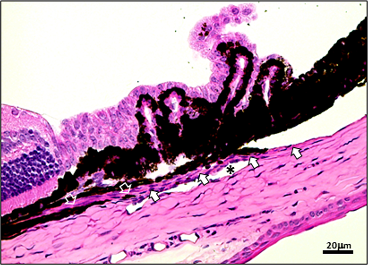
 Figure 1 of
Ko, Mol Vis 2013; 19:2561-2570.
Figure 1 of
Ko, Mol Vis 2013; 19:2561-2570.