Figure 3. Hematoxylin and Eosin (H and E), and transmission electron microscopy (TEM) of the engrafted human conjunctival epithelia
(CjE). A, B: H and E stain of corneas with or without transplanted epithelial sheets two weeks after transplantation. C, D: TEM of the grafted tissue showing hemidesmosomal profiles at the basal cell-central corneal stromal interface (C, arrows) and desmosomes between wing shaped cells (D, arrowheads). Representative images shown of four grafted corneas.
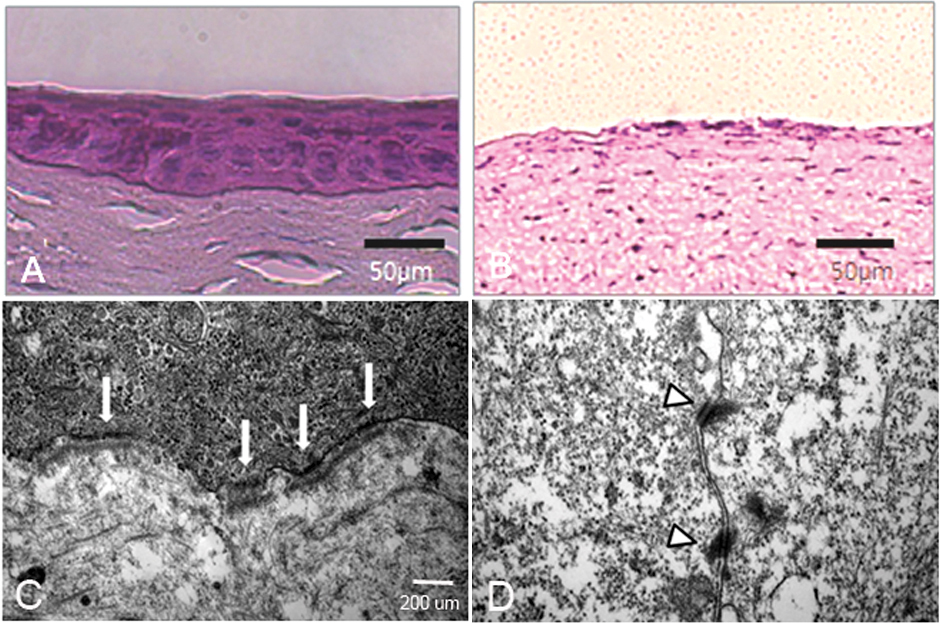
 Figure 3 of
Jeon, Mol Vis 2013; 19:2542-2550.
Figure 3 of
Jeon, Mol Vis 2013; 19:2542-2550.