Figure 9. Ephrin-A5 expression is observed in several parts of the developing eye. A and E: EphA5-AP staining is observed in the lens epithelium (le), lens bow (lb), cornea (c), and ciliary body (cb) in the E14 wild-type
eye while absent in the ephrin-A5-/- animal. B and F: Expression of ephrin-A ligands are maintained in the wild-type at P0 though in lower levels in comparison with earlier embryonic
stages, while remaining absent in the ephrin-A5-/- eye. C and G: Ephrin-A ligand expression is observed in the lens bow region of P7 wild-type mice while absent in ephrin-A5-/- mice at the same age. D and H: High levels of ephrin-A ligand is observed in the cornea of wild-type mice at P7 and not present in ephrin-A5-/- mice at the same age. Scale bars are in micrometers.
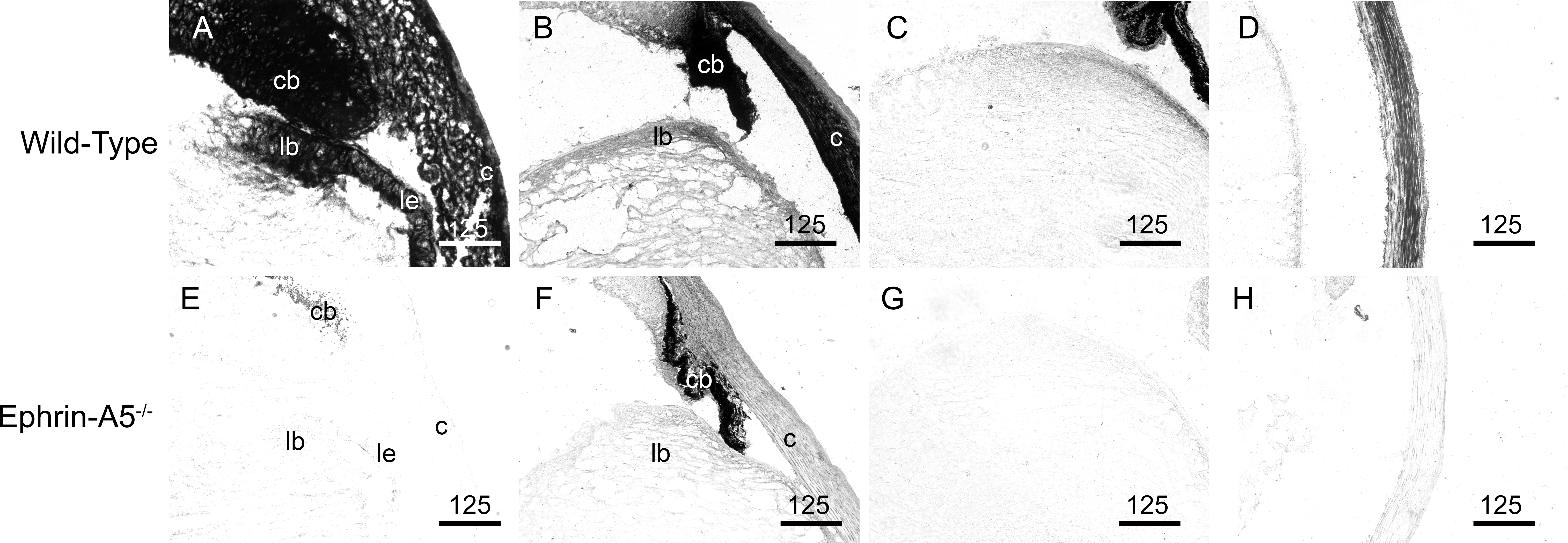
 Figure 9 of
Son, Mol Vis 2013; 19:254-266.
Figure 9 of
Son, Mol Vis 2013; 19:254-266.