Figure 4. Distinct alterations in cell shape are observed in the ephrin-A5-/- lens fiber cell layers. A-F: Wild-type (A-C) and ephrin-A5-/- (D-F) P21 lenses are labeled for ZO-1 (A and D) and β-Catenin (B and E) to delineate cell borders or to distinguish distinct lens fiber areas. Disruptions in fiber cell organization are observed
in the ephrin-A5-/- lens. Scale bar = 100 μm. G-I: Disorganization of the fiber cells in the ephrin-A5-/- lens are observed in all fiber cell regions, including the cortical (G), subcortical (H), and central (I) areas. Scale bar=50 μm.
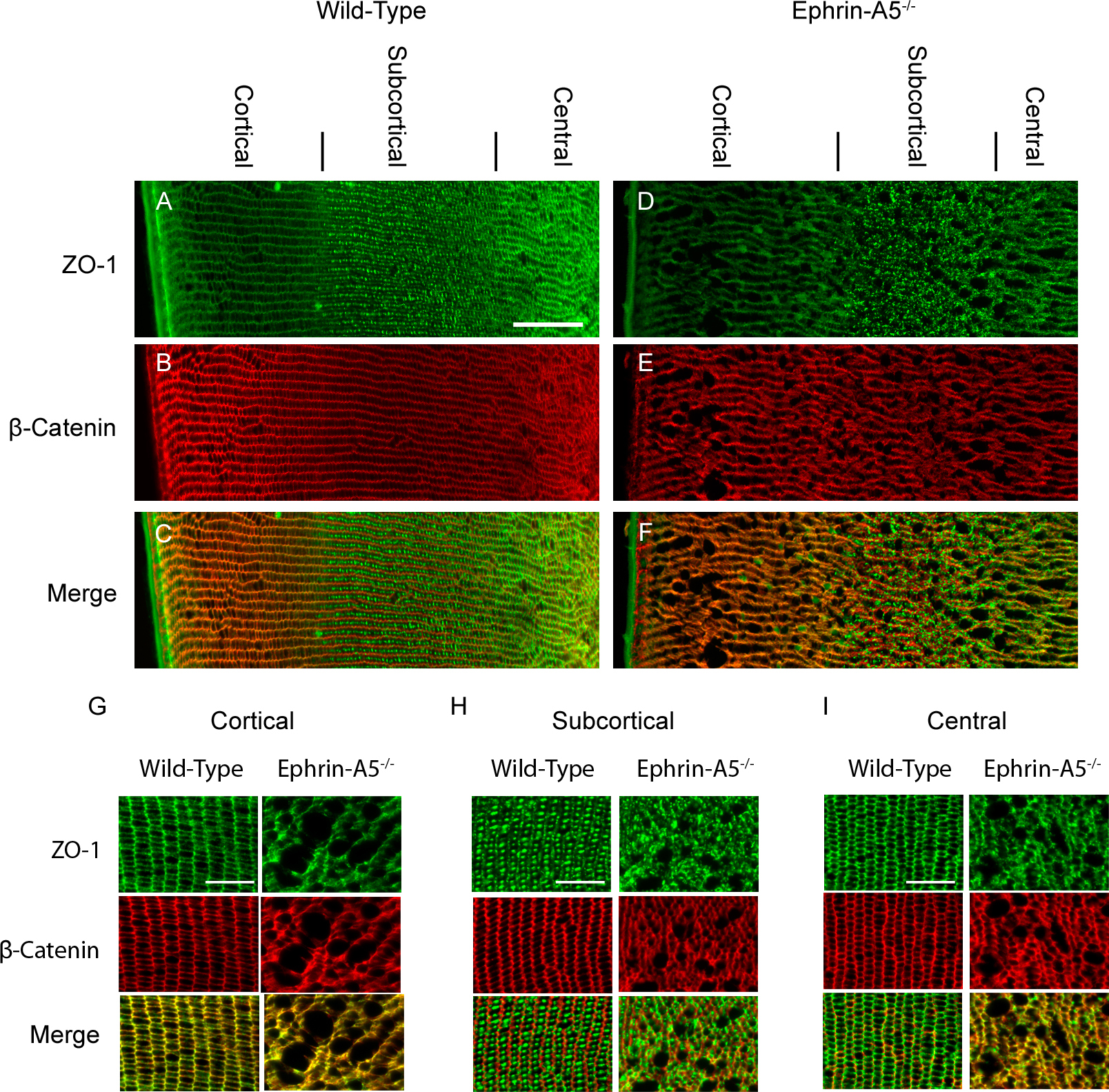
 Figure 4 of
Son, Mol Vis 2013; 19:254-266.
Figure 4 of
Son, Mol Vis 2013; 19:254-266.