Figure 3. Deformations in the structure of ephrin-A5-/- lenses occur in postnatal eyes. A-F: Embryonic development of wild-type (WT; A-C) and ephrin-A5-/- (D-F) lenses appear similar, with no abnormalities observed in the early ephrin-A5-/- lens. Scale bars in mm. G-L: While wild-type lenses (G-I) show no deformities in postnatal stages, ephrin-A5-/- lenses (J-L) display noticeable lens deficits by P21 with the presence of vacuoles around the lens bow (compare H and K, see arrows). The deficits become progressively more severe, as larger vacuoles and complete posterior lens rupture is observed
by P60 (Compare I and L, see arrow). Scale bars in mm.
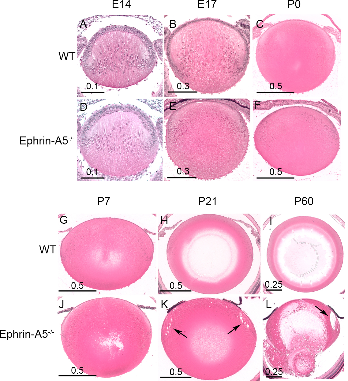
 Figure 3 of
Son, Mol Vis 2013; 19:254-266.
Figure 3 of
Son, Mol Vis 2013; 19:254-266.