Figure 5. Colocalization of inhibitor of apoptosis and X-linked inhibitor of apoptosis proteins with neurofilament H (a neuronal marker)
and glial fibrillary acidic protein (a glial marker). Triple immunostaining of X-linked inhibitor of apoptosis (XIAP) and
IAP with glial fibrillary acidic protein (GFAP) and neurofilament H (NFH) was performed on optic nerves (ONs) at 8 days after
the induction of elevated intraocular protein (IOP). A: Colocalization of IAP-1 with NFH and GFAP suggests that IAP-1 in the ON is mostly neuronal. B: Colocalization of XIAP with NFH and GFAP suggests that XIAP in the ON is mostly glial. C, D: These images represent colocalization of IAP-1 and XIAP with NFH and GFAP (white areas). The analysis was done after the
pictures were deconvoluted using Hyugens program with the Imaris Bitmate, as described in Material and Methods; n=5 for each
staining, *p<0.05, **p<0.001.
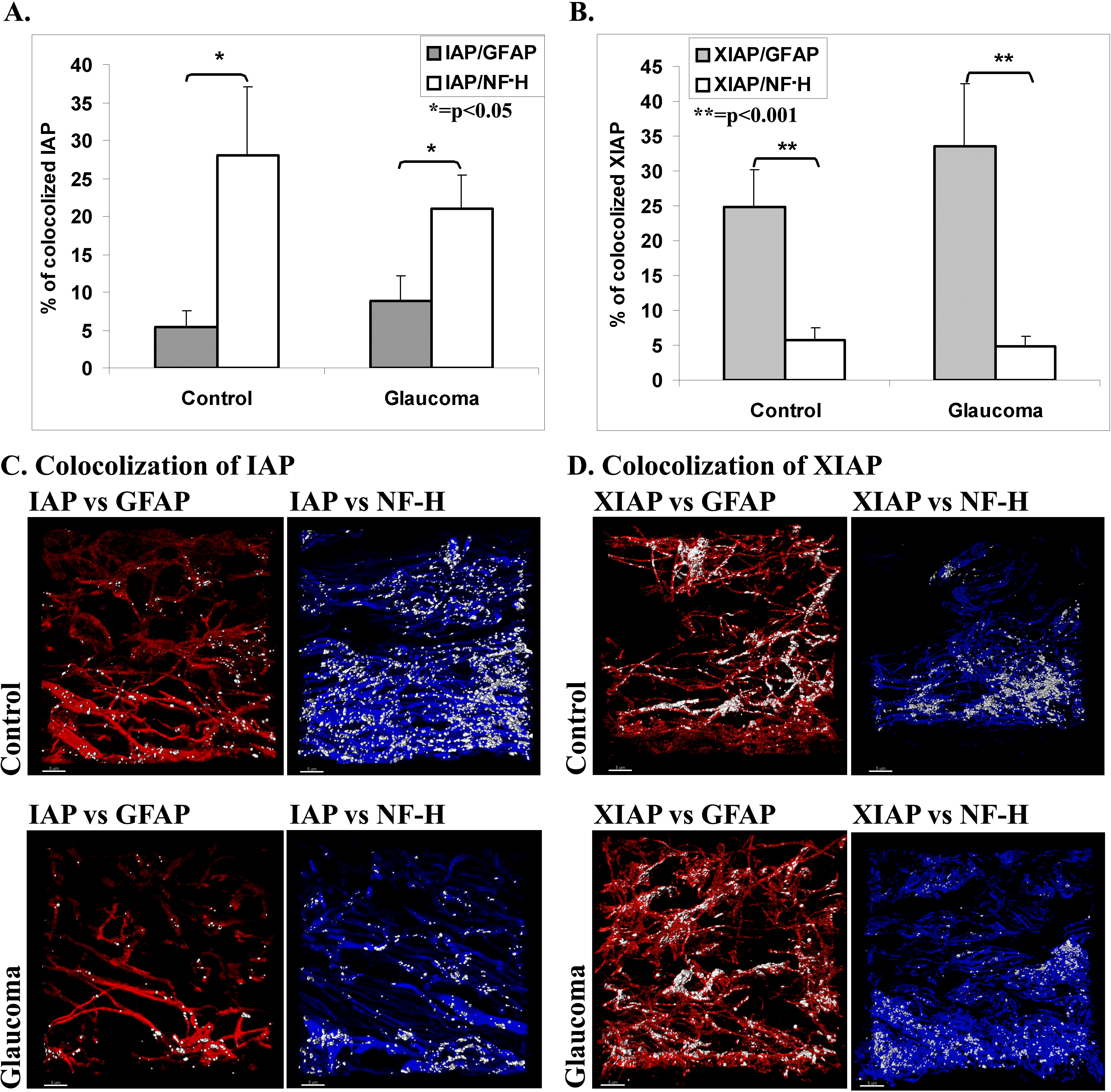
 Figure 5 of
Levkovitch-Verbin, Mol Vis 2013; 19:2526-2541.
Figure 5 of
Levkovitch-Verbin, Mol Vis 2013; 19:2526-2541.