Figure 3. X-linked inhibitor of apoptosis and inhibitor of apoptosis-1 protein expressions in the optic nerves of glaucomatous eyes
and their controls. A and B: Colocalization of X-linked inhibitor of apoptosis (XIAP) and IAP-1 with glial fibrillary acidic protein (GFAP; a glial marker)
and neurofilament-H (NFH, a neuronal marker) at 8 days after the induction of elevated intraocular pressure (IOP) suggests
that both XIAP and IAP-1 are present in optic nerve (ON) axons and glia. IAP-1 was more colocalized with NFH and XIAP was
more colocalized with GFAP.
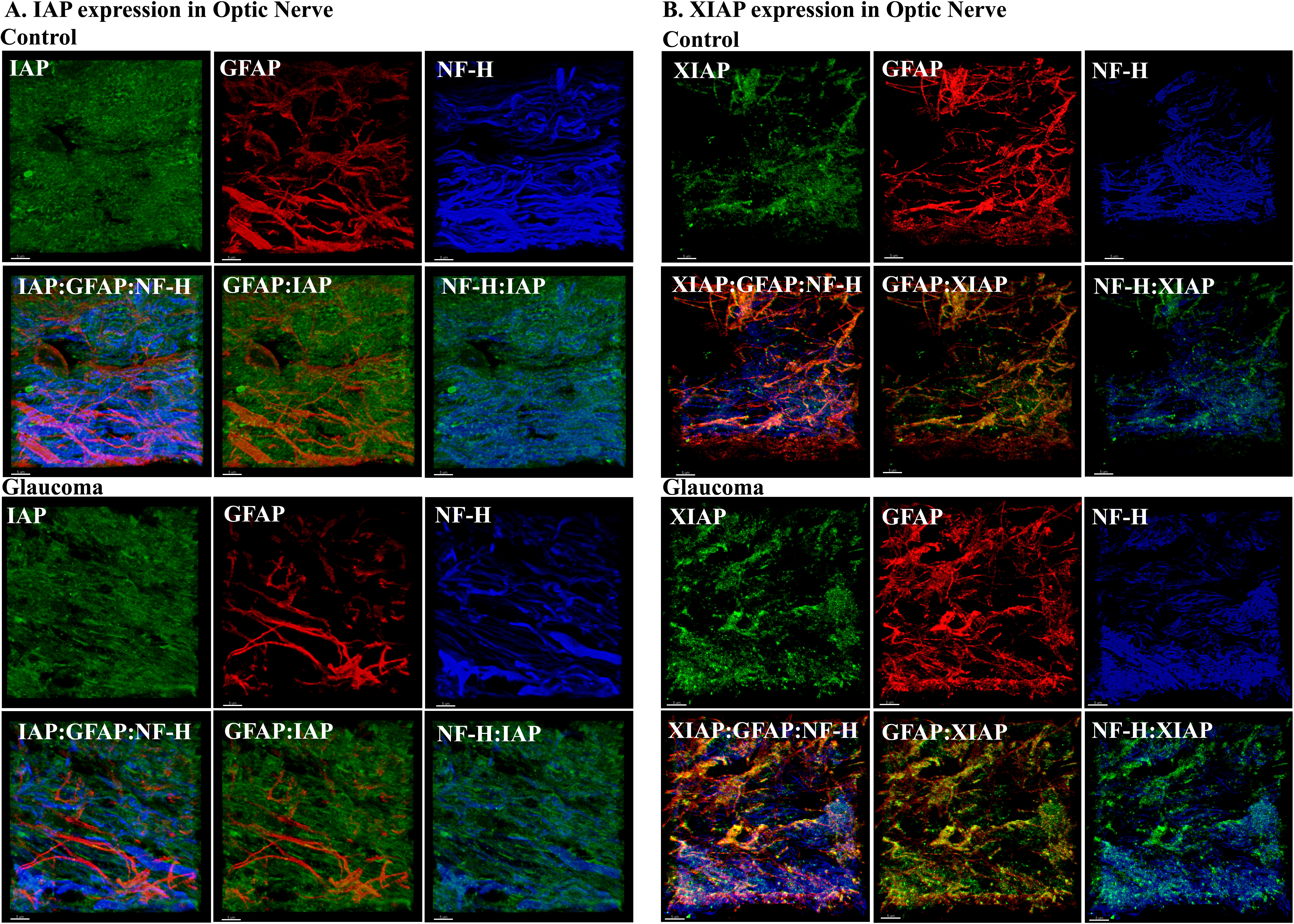
 Figure 3 of
Levkovitch-Verbin, Mol Vis 2013; 19:2526-2541.
Figure 3 of
Levkovitch-Verbin, Mol Vis 2013; 19:2526-2541.