Figure 2. X-linked inhibitor of apoptosis (XIAP) and inhibitor of apoptosis-1 (IAP-1) protein expressions increased in retinas of glaucomatous
eyes compared to controls. A and B: Both were expressed in the retinal ganglion cell (RGC) layer and in other retinal layers. Colocalization with glial fibrillary
acidic protein (GFAP; a glial marker) and Thy-1 (an RGC marker) at 8 days after the induction of elevated intraocular pressure
(IOP) suggests that both inhibitor of apoptosis (IAP)-1 and X-linked IAP (XIAP) secretion in the retina originate from both
RGC bodies and glial cells.
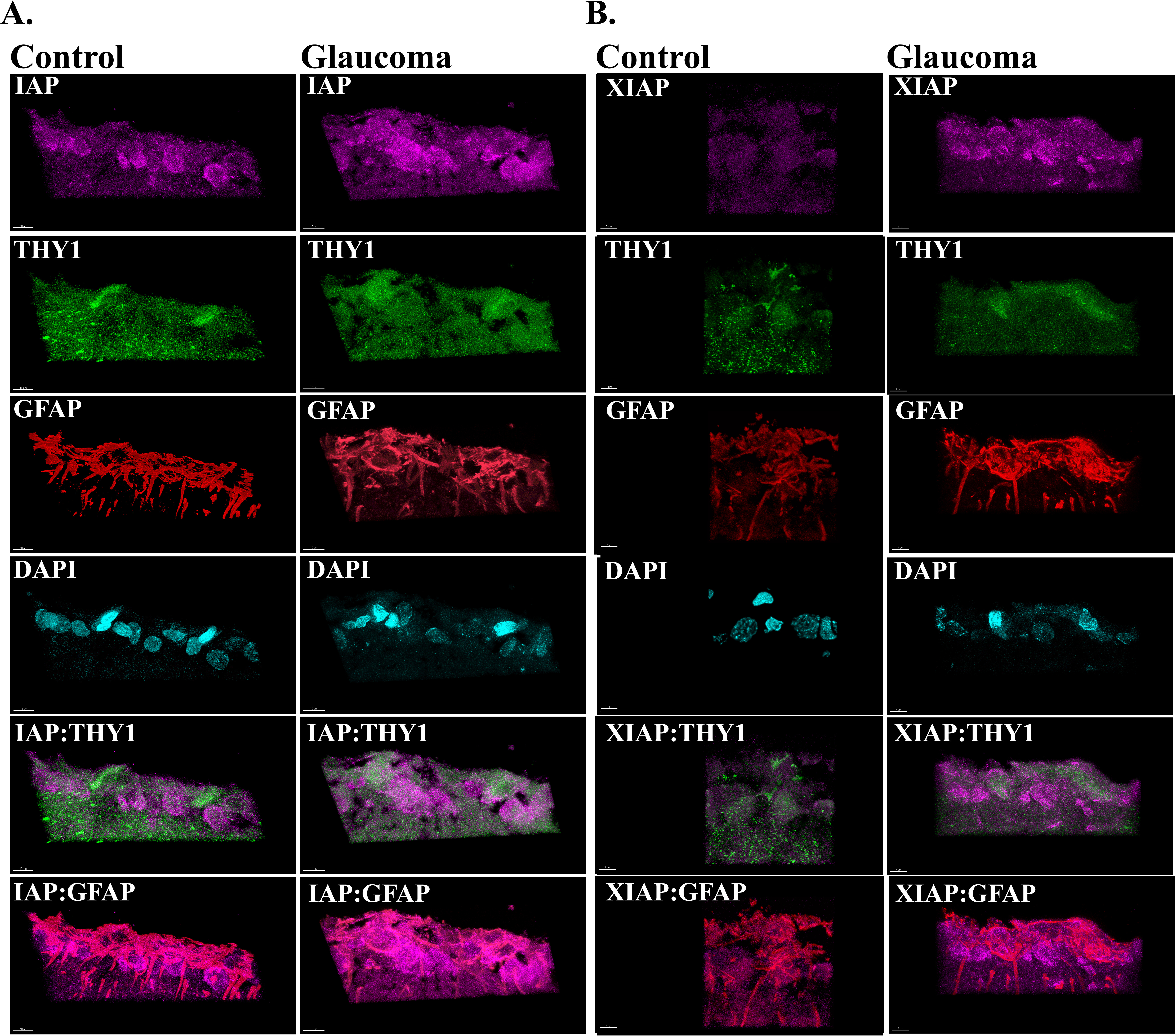
 Figure 2 of
Levkovitch-Verbin, Mol Vis 2013; 19:2526-2541.
Figure 2 of
Levkovitch-Verbin, Mol Vis 2013; 19:2526-2541.