Figure 4. A larger number of dendritic cells infiltrate the cornea of D6−/− mice following inflammation induced by cauterization. A: Confocal microscopy of the cornea from wild-type (WT; D6+/+) and D6−/− mice 7 days post-cornea cauterization. The cornea was stained for CD11c (green) cells (magnification 40X). B: The bar chart represents the quantification of the CD11c-infiltrated cells in the D6−/− and WT corneas 7 days post-cornea cauterization. *p≤0.05.
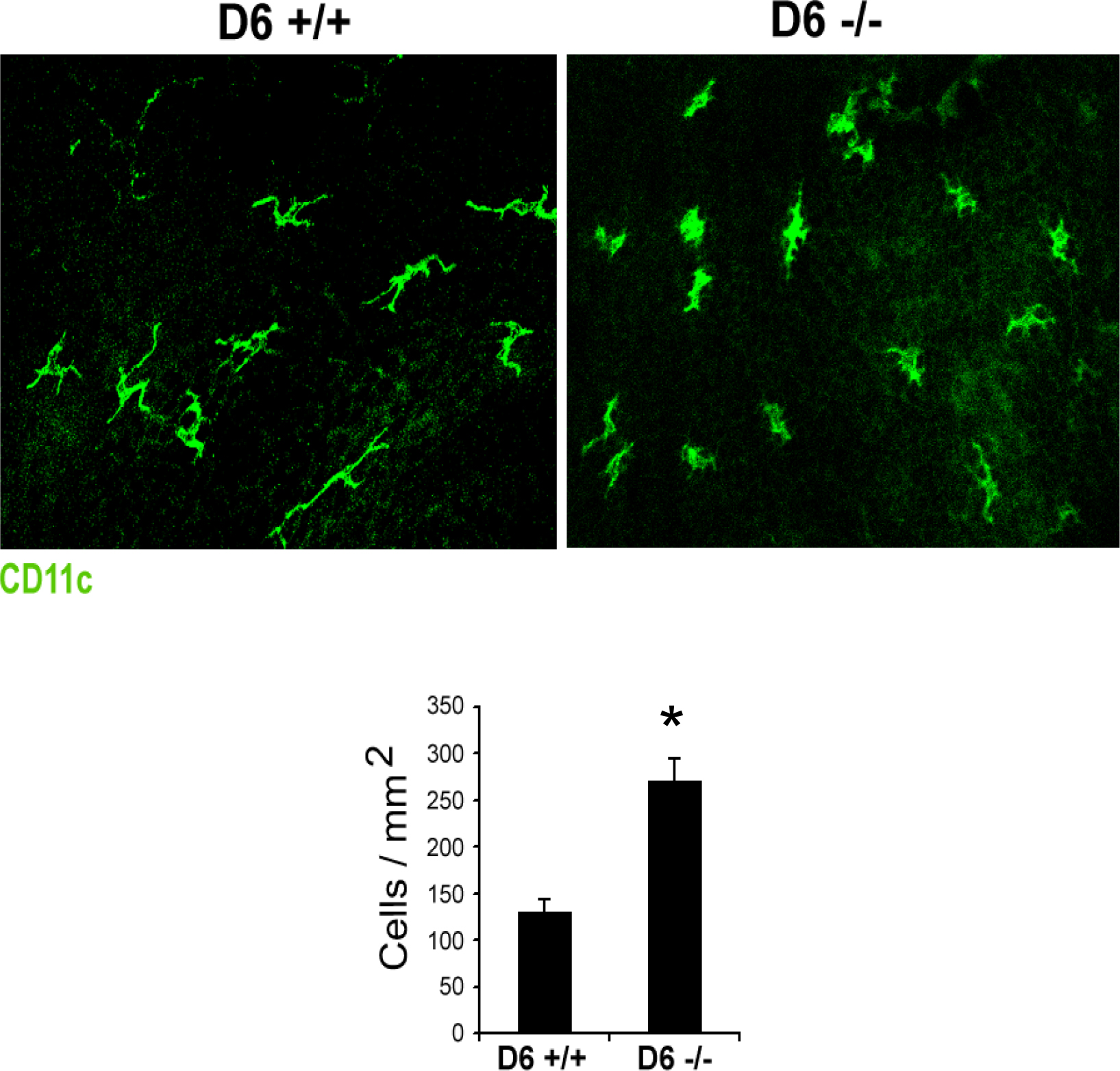
 Figure 4 of
Hajrasouliha, Mol Vis 2013; 19:2517-2525.
Figure 4 of
Hajrasouliha, Mol Vis 2013; 19:2517-2525.