Figure 1. Primary corneal tissue culture. This figure represents primary tissue culture from sample no. 4 under light microscopy. A: The migration of cells out of the tissue started on day 7 after seeding (white arrow shows margin of tissue, 10X magnification).
B: One of the cell colonies was performed after 15 days post-seeding (20X magnification). C, D: After cell culture expansion, the cultivated cells were starved for 24 h to compare the morphological difference between
fibroblasts and keratocytes (C, D, respectively).
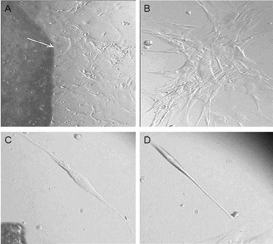
 Figure 1 of
Saee-Rad, Mol Vis 2013; 19:2501-2507.
Figure 1 of
Saee-Rad, Mol Vis 2013; 19:2501-2507.