Figure 2. Clinical features of patients with mutations in autosomal recessive retinitis pigmentosa. A-F: Family RP1361 with PDE6A mutation; fundus photographs of right eyes of subjects II:1 (A) and II:2 (B), retinal autofluorescence in right eyes of subjects II:1 (C) and II:2 (D), OCT scan of the macula of the right eye of subjects II:1 (E) and II:2 (F). G-H: Family RP1324 with CNGB1 mutation; fundus photographs of right eyes of subjects II:8 (G) and II:3 (H). I-J: Family RP1013 with C2ORF71 mutation; fundus photograph (I) and retinal autofluorescence (J) of the left eye of subject II:2.
K-P: Families RP1625 (K-N) and RP854 (O, P) with RP1 mutations; fundus photograph (K) and retinal autofluorescence (L) of the right eye of subject II:2 of family RP1625, OCT scan in the macula of the right
(M) and left (N) eyes of subject II:2 of family RP1625, fundus photographs of the left eye of subject II:2 of family RP854
showing the macula (O) and the temporal periphery (P). Q-T: Families RP517 (Q-S) and RP1682 (T) with RLBP1 mutations; fundus photographs of the right eye at 32 years (Q) and 40 years (R) and of the left
eye at 40 years (S) of subject II:1 of family RP517, and of the left eye (T) of subject II:3 of family RP1682.
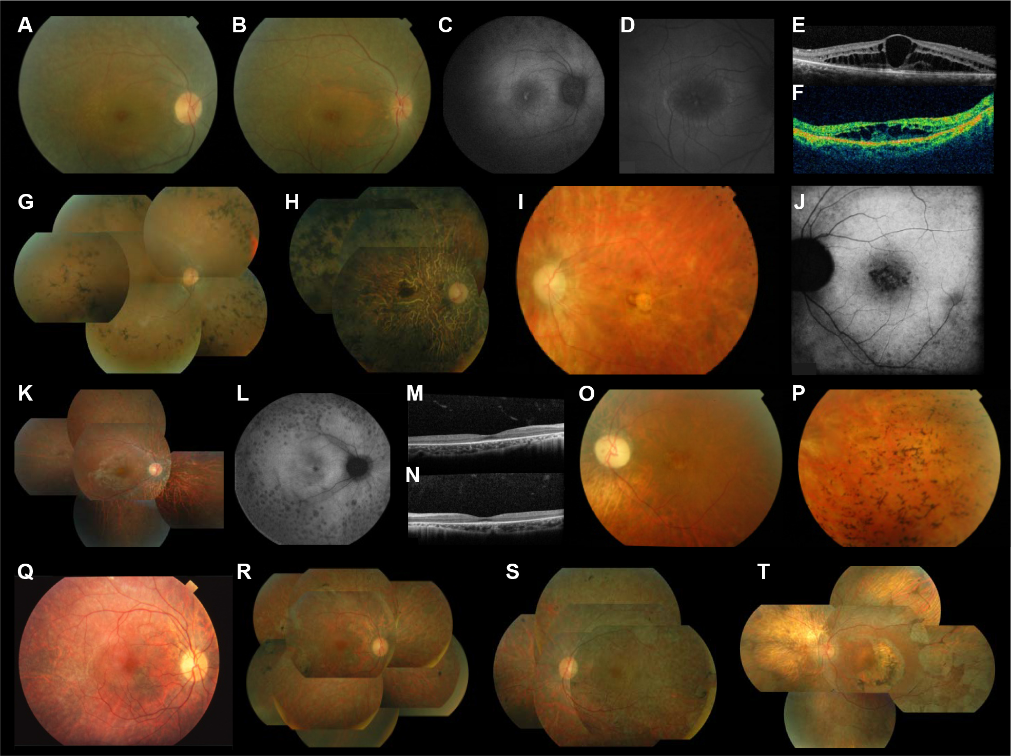
 Figure 2 of
Bocquet, Mol Vis 2013; 19:2487-2500.
Figure 2 of
Bocquet, Mol Vis 2013; 19:2487-2500.