Figure 5. Immunohistochemical staining showed that the intensity of MMP-10 staining (upper panel) was increased by exposure to 50% galactose,
while integrin α3 expression (lower panel) decreased. These changes were normalized with treatment of aldose reductase inhibitor.
The scale bar indicates 50 μm.
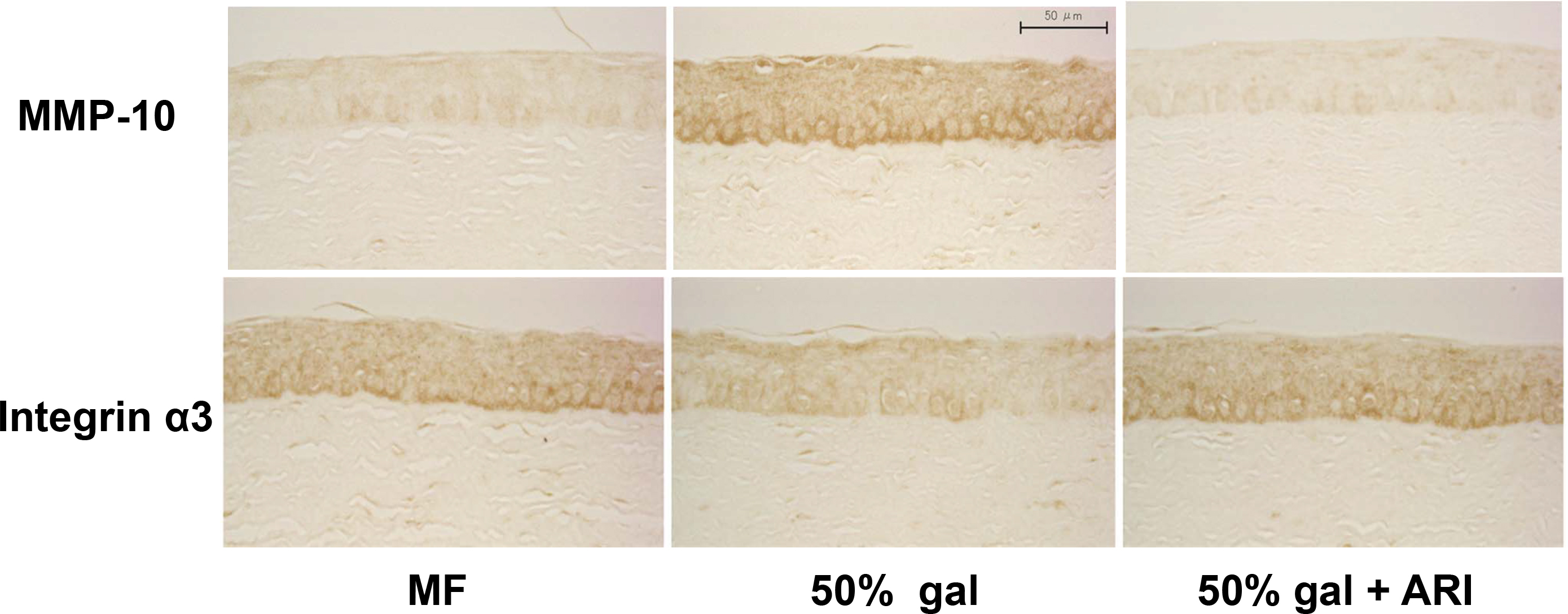
 Figure 5 of
Takamura, Mol Vis 2013; 19:2477-2486.
Figure 5 of
Takamura, Mol Vis 2013; 19:2477-2486.