Figure 2. Light microscopy of lenses of newborn and 1-month-old wild-type and human p53 homozygous transgenic (Tgp53t/t) mice. A: Newborn wild-type mice show an external capsule (black arrow); epithelial cells line the front surface of the organ and
at the equatorial region (arrow head) transform into highly elongated, ribbon-like fiber cells (white arrow). B: In newborn Tgp53t/t mice, the capsule (black arrow) and the superficial epithelial cells (arrow head) are also visible, but some structural disorganization
can already be observed in the interior (white arrow). C: In 1-month-old wild-type mice, the capsule (black arrow), the single layer of epithelial cells at the front surface (arrow
head), and the fiber cells organized in layers (white arrow) are observed. D: In 1-month-old Tgp53t/t mice, the capsule (black arrow) is still visible, but the superficial epithelial cells are not present, and disorganization
is increased in the interior of the organ (white arrow). Bar: 20 µm.
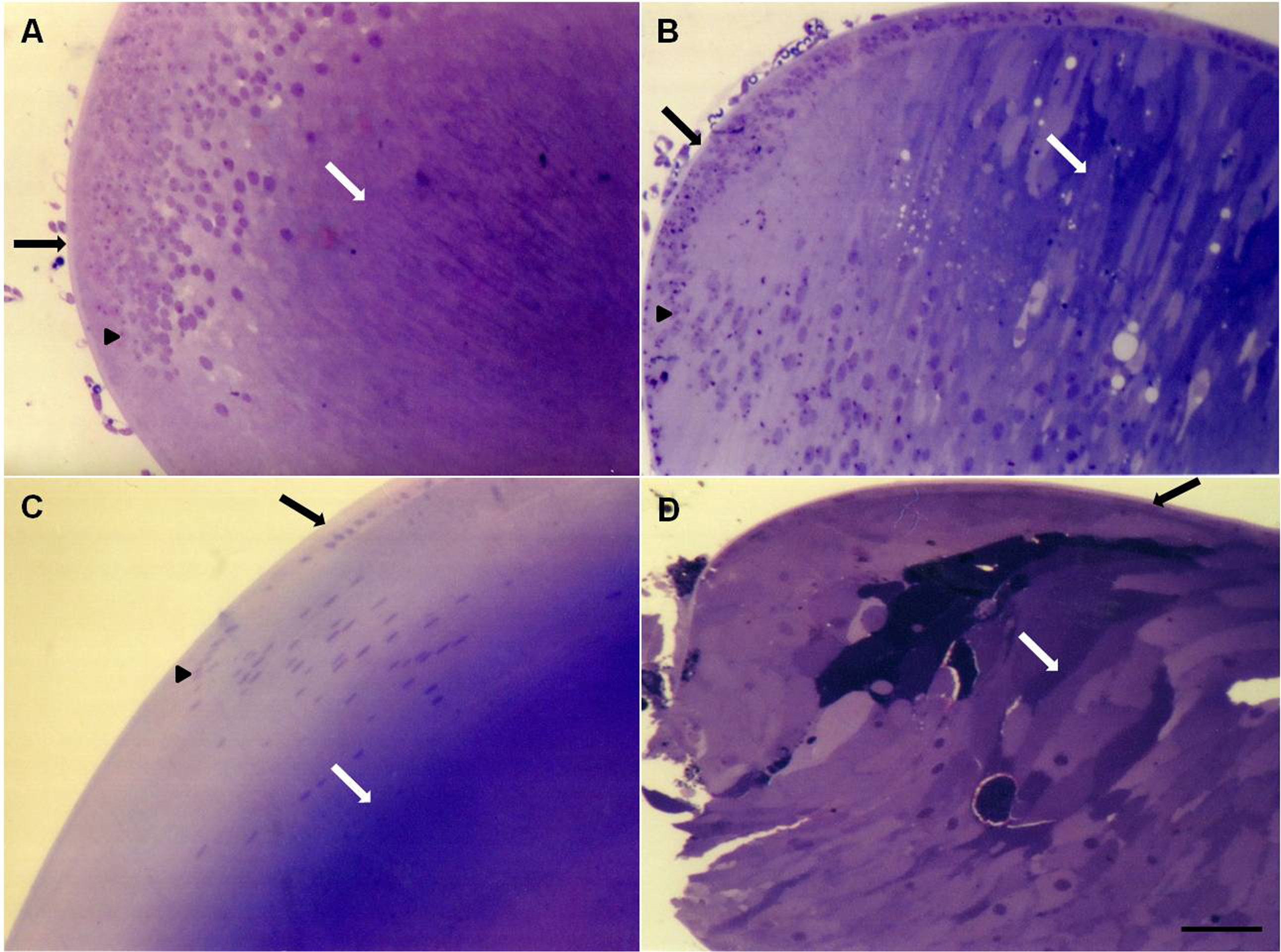
 Figure 2 of
Jaramillo-Rangel, Mol Vis 2013; 19:2468-2476.
Figure 2 of
Jaramillo-Rangel, Mol Vis 2013; 19:2468-2476.