Figure 1. Light and electron microscopy of lenses from 2-month-old wild-type and human p53 homozygous transgenic (Tgp53t/t) mice. A: Wild-type mice show by light microscopy an extracellular collagenous capsule (black arrow), a surface monolayer of epithelial
cells (arrow head), and elongated fiber cells (white arrow). Bar: 20 µm. B: Analysis of Tgp53t/t mice by light microscopy demonstrate that the external capsule is not visible, the nuclei of the apoptotic cells form superficial
clusters (black arrow), and the interior of the organ has a highly disordered structure and nuclei with condensed chromatin
(white arrow). Bar: 20 µm. C: Wild-type mice show by electron microscopy a collagenous capsule (asterisk), epithelial cells with nuclei with normal morphology
(black arrow), and fiber cells organized in layers (white arrow). Bar: 5 µm. D: In the electron microscopy analysis of lenses of Tgp53t/t mice, the interior does not show a defined order or evidence of normal fiber cells. Apoptotic bodies (arrows) are visible.
Bar: 1 µm.
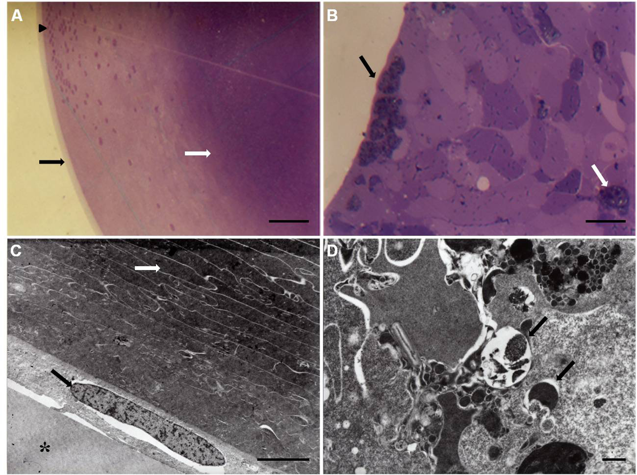
 Figure 1 of
Jaramillo-Rangel, Mol Vis 2013; 19:2468-2476.
Figure 1 of
Jaramillo-Rangel, Mol Vis 2013; 19:2468-2476.