Figure 9. JC-1 analysis of bovine lens epithelial cells treated with UO126 inhibitor. Secondary cultures of bovine lens epithelial cells
were incubated for 90 min with serum-free minimal essential media (MEM), under atmospheric condition, containing 10 µM UO126
or 0.05% DMSO vehicle. Cells were switched to hypoxia for 3h in the continued presence or absence of UO126. At the end of
the hypoxic exposure, the cell media were removed, and fresh, oxygenated serum-free MEM containing 5 µg/ml JC-1 and either
UO126 or DMSO added for 30 min in atmospheric oxygen. At the end of the 30 min incubation period, the media were again switched
to fresh serum-free MEM containing UO126 or DMSO in the absence of the JC-1 dye. A random field of cells was chosen, and that
field of cells was imaged every 150 s for 60 min. Serial confocal imaging of mitochondrial depolarization of the secondary
cultures of normal bovine lens epithelial cells in the presence of UO126 demonstrated significant depolarization compared
to control cells (top panel). Images of the red and green intensity (bottom panel) for the UO126- and DMSO-treated cells at
t=60 min (bar=20 µm) are shown (bottom panel). Note the marked intensity of the green channel with the UO126-treated cells
relative to DMSO mock treatment at the completion of the 60 min analysis, which indicates a propensity toward a significant
degree of mitochondrial depolarization.
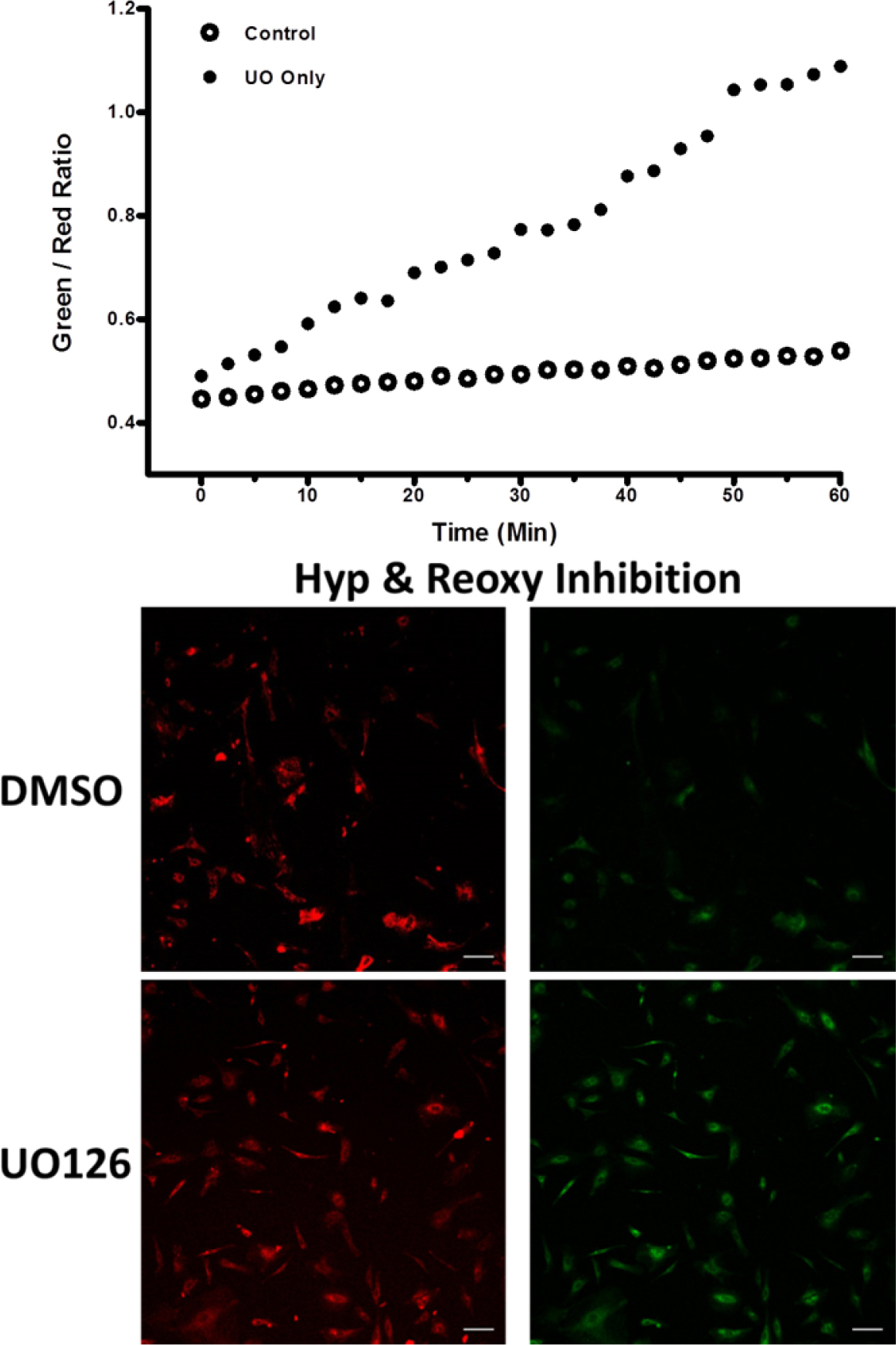
 Figure 9 of
Brooks, Mol Vis 2013; 19:2451-2467.
Figure 9 of
Brooks, Mol Vis 2013; 19:2451-2467.