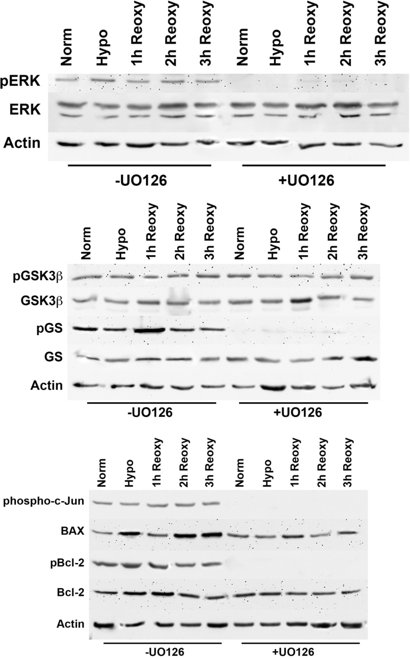
Figure 8. Western blot analysis of GSK-3β and GS phosphorylation and BAX, Bcl-2, pBcl-2, and phospho-c-Jun in secondary cultures of
normal bovine cells treated with UO126 inhibitor. Bovine cell cultures were incubated for 90 min in serum-free minimal essential
media (MEM), under conditions of atmospheric oxygen, containing either 10 µM UO126 or 0.05% DMSO) vehicle. Cells were then
exposed to hypoxia for 3 h in the presence of either UO126 or DMSO vehicle. At the end of the hypoxic incubation period, the
hypoxic media were removed, and fresh, oxygenated serum-free MEM with UO126 or DMSO were added to the cultures. Cells were
then placed in atmospheric oxygen for up to 3 h. Cultures were collected after (1) continuous normoxic exposure (about 21%
oxygen), (2) after the 3 h hypoxic exposure (about 1% oxygen), or (3) after reintroduction of atmospheric oxygen (about 21%)
for 1, 2, or 3 h subsequent to the 3 h hypoxic exposure. Total cell lysates were analyzed with immunoblots using 25 µg of
protein per lane. Anti-actin was used to normalize the bands to ensure equivalent lane loading. Prevention of phosphorylation
of ERK with UO126 treatment was noted (top panel), as was the inhibition of phosphorylated glycogen synthase with western
blot analysis (middle panel). The levels of GSK-3β) and pGSK-3β were consistent in the presence and absence of UO126. The
phosphorylation of c-Jun, as well as that of Bcl-2, was blocked by treatment with UO126 under all conditions as determined
with western blot analysis (bottom panel). Of particular note, Bcl-2 levels were unaffected by UO126 treatment compared to
HLE-B3 cells (refer to
Figure 5) where a significant diminution of Bcl-2 was observed but only upon reintroduction to atmospheric oxygen. No change in the
relative levels of BAX was evident by treatment with UO126 under any condition. The experiment with normal, secondary cultures
of bovine cells was run once, to confirm that a similar pattern of biochemical modifications by treatment with UO126 was reproducible
with normal bovine cell cultures as was observed with HLE-B3, affirming that the former results were not influenced by viral
transformation.
 Figure 8 of
Brooks, Mol Vis 2013; 19:2451-2467.
Figure 8 of
Brooks, Mol Vis 2013; 19:2451-2467.