Figure 4. Western blot analysis of BAX, Bcl-2, pBcl-2, and phospho-c-Jun in HLE-B3 cells in the presence or absence of SB216763. Total
cell lysates were collected from >85% confluent HLE-B3 cell cultures that were incubated for 90 min in serum-free minimal
essential media (MEM), under conditions of atmospheric oxygen, containing either 12 µM SB216763) or 0.05% DMSO vehicle. Cells
were then exposed to hypoxia for 3 h in the continued presence of SB216763 or DMSO vehicle. At the end of the hypoxic incubation
period, the hypoxic media were removed, and fresh, oxygenated serum-free MEM with SB216763 or DMSO were added to the cultures.
Cells were then placed in atmospheric oxygen for up to 3 h. Cultures were collected after (1) continuous normoxic exposure
(about 21% oxygen), (2) after the 3 h hypoxic exposure (about 1% oxygen), or (3) after reintroduction of atmospheric oxygen
(about 21%) for 1, 2, or 3 h subsequent to the 3 h hypoxic exposure. Total cell lysates were analyzed with immunoblots using
25 µg of protein per lane. Anti-actin was used to normalize the bands to ensure equivalent lane loading. Three experiments,
using independent cell populations, were quantified using GraphPad Prism 5 and the relative densities plotted for BAX/actin,
Bcl-2/actin, pBcl-2/actin, and phospho-c-Jun/Actin. No change was evident in the ratio of BAX/actin, Bcl-2/actin, pBcl-2/actin,
or phospho-c-Jun/actin by treatment with SB216763. Error bars represent standard error, Student t test.
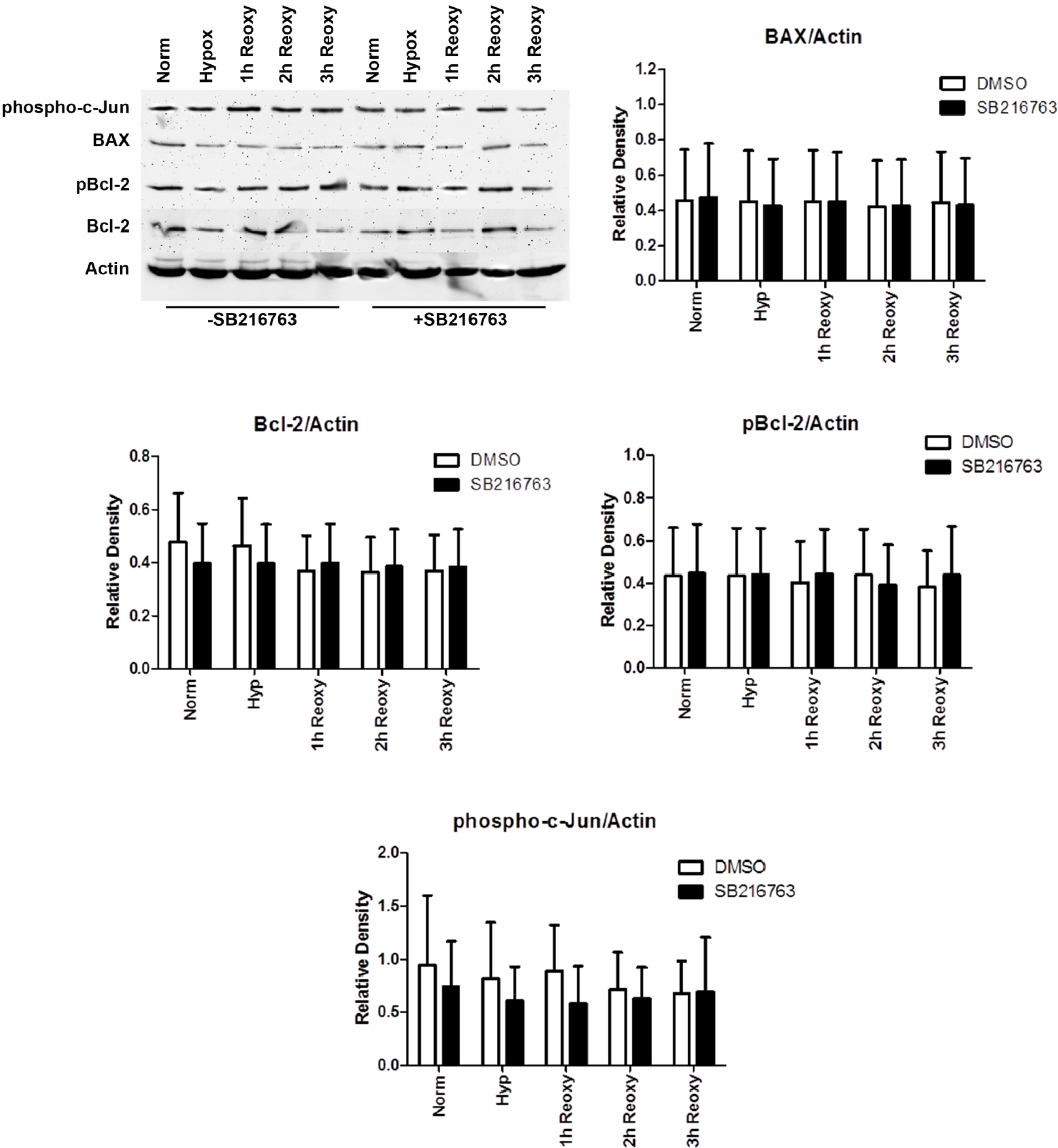
 Figure 4 of
Brooks, Mol Vis 2013; 19:2451-2467.
Figure 4 of
Brooks, Mol Vis 2013; 19:2451-2467.