Figure 3. Western blot analysis of GSK-3β and GS phosphorylation in HLE-B3 cells treated with UO126 inhibitor. HLE-B3 cell cultures
were incubated for 90 min in serum-free minimal essential media (MEM), under conditions of atmospheric oxygen, containing
either 10 µM UO126 or 0.05% DMSO vehicle. Cells were then exposed to hypoxia for 3 h. At the end of the hypoxic incubation
period, the hypoxic media were removed, and fresh, oxygenated serum-free MEM with UO126 or DMSO vehicle were added to the
cultures. Cells were then placed in atmospheric oxygen for up to 3 h. Cultures were collected after (1) continuous normoxic
exposure (about 21% oxygen), (2) after the 3 h hypoxic exposure (about 1% oxygen), or (3) after reintroduction of atmospheric
oxygen (about 21%) for 1, 2, or 3 h subsequent to the 3 h hypoxic exposure. Total cell lysates were analyzed with immunoblots
using 25 µg of protein per lane. Anti actin was used to normalize the bands to ensure equivalent lane loading. The loss of
phosphorylation of ERK by UO126 treatment was noted (top, left panel), as was the loss of phosphorylated glycogen synthase
by western blot analysis (top, right panel). Three experiments, using independent cell populations, were quantified using
GraphPad Prism 5, and the relative densities were plotted for pGSK-3β/GSK-3β and pGS/GS. No change was evident in the ratio
of pGSK-3β/GSK-3β while significant inhibition of the phosphorylation of glycogen synthase by UO126 was indicated (middle
panel). Error bars represent standard error. The asterisks (***) indicate p<0.001, Student t test. (bottom panel, left) HLE-B3 cells were incubated for 90 min with serum-free MEM, under atmospheric condition, containing
10 µM UO126 or 0.05% DMSO vehicle. Cells were switched to hypoxia for 3 h in the continued presence of UO126 or DMSO vehicle.
At the end of the hypoxic exposure, the cells had their media removed, and fresh, oxygenated serum-free MEM containing 5 µg/ml
JC-1 and either UO126 or DMSO added for 30 min in atmospheric oxygen. At the end of the 30 min incubation period, the media
were again switched with fresh serum-free MEM containing UO126 or DMSO in the absence of the JC-1 dye. The same field of cells
was imaged every 150 s for 60 min. Serial confocal imaging of mitochondrial depolarization in HLE-B3 cells in the presence
of UO126 demonstrated significant depolarization compared to control cells. (bottom panel, left) Images of the red and green
intensity for the UO126- and DMSO-treated cells at t=60 min (bar=20 µm). Note the marked intensity of the green channel with
UO126-treated cells relative to DMSO mock treatment at the completion of the 60 min analysis (bottom panel, right) indicating
mitochondrial depolarization.
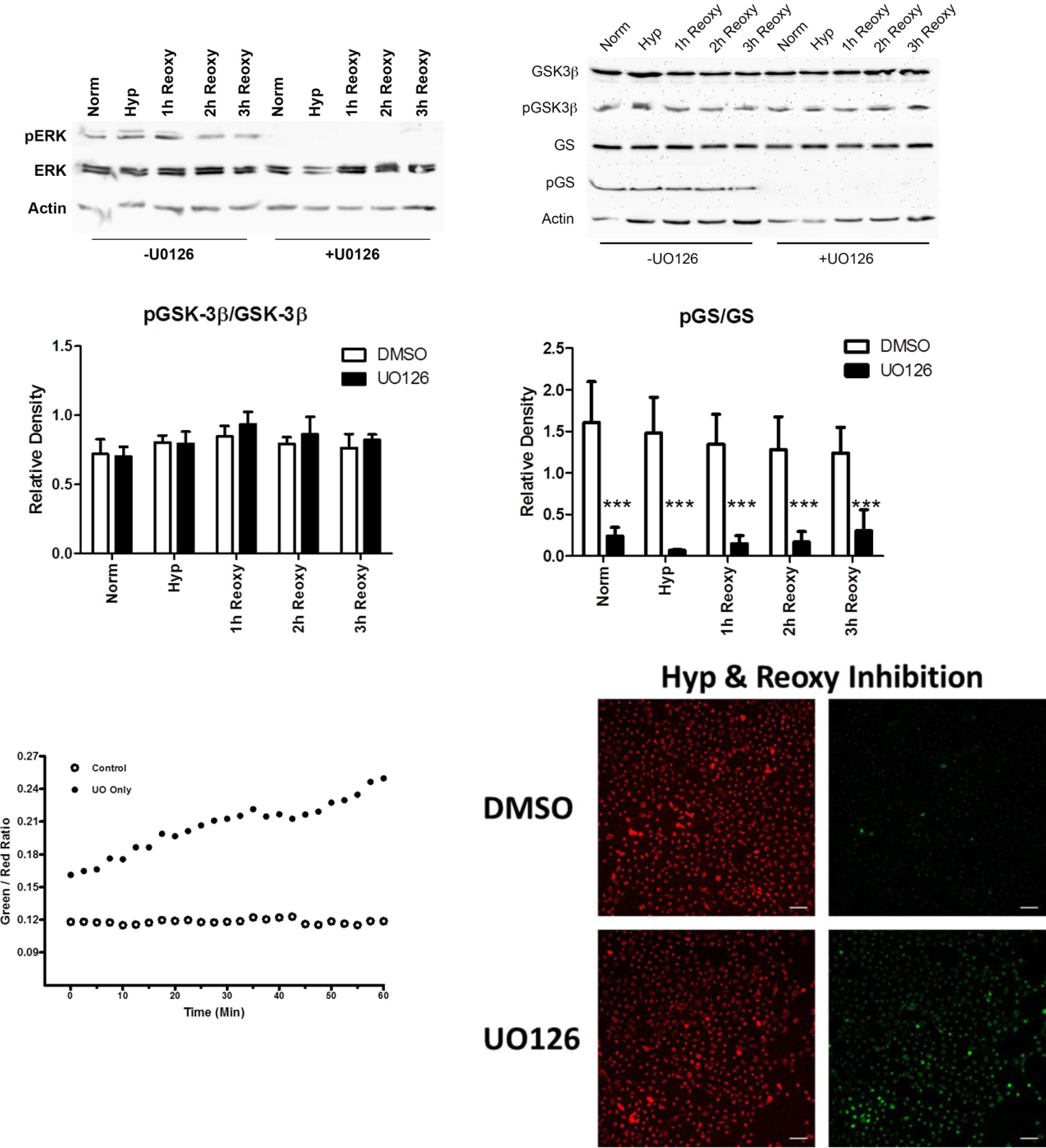
 Figure 3 of
Brooks, Mol Vis 2013; 19:2451-2467.
Figure 3 of
Brooks, Mol Vis 2013; 19:2451-2467.