Figure 7 of
Kumar, Mol Vis 2013; 19:2436-2450.
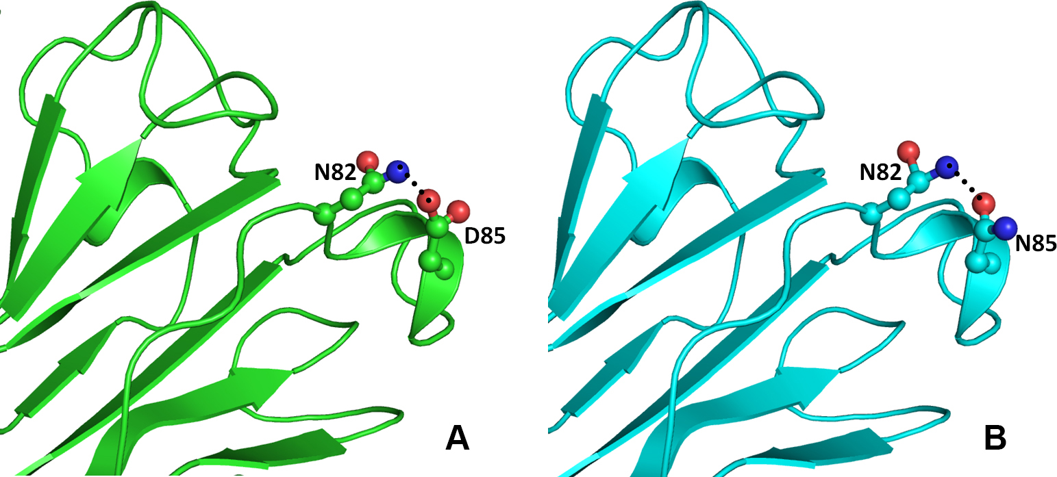
Figure 7.
Model structure representation of the wild and mutant (Asp85Asn) proteins. In both structures (
A
and
B
), the interaction of residues as balls and sticks and hydrogen bonds as black dotted lines in beta crystallin B1 protein is same.
 Figure 7 of
Kumar, Mol Vis 2013; 19:2436-2450.
Figure 7 of
Kumar, Mol Vis 2013; 19:2436-2450.