Figure 6. Model structure representation of the wild and mutant (Glu75Lys) proteins. A: Beta crystallin B1 (CRYBB1) protein showing the important residues (balls and sticks) and the hydrogen bonds (black dotted lines). B: The contacts are lost in the mutant.
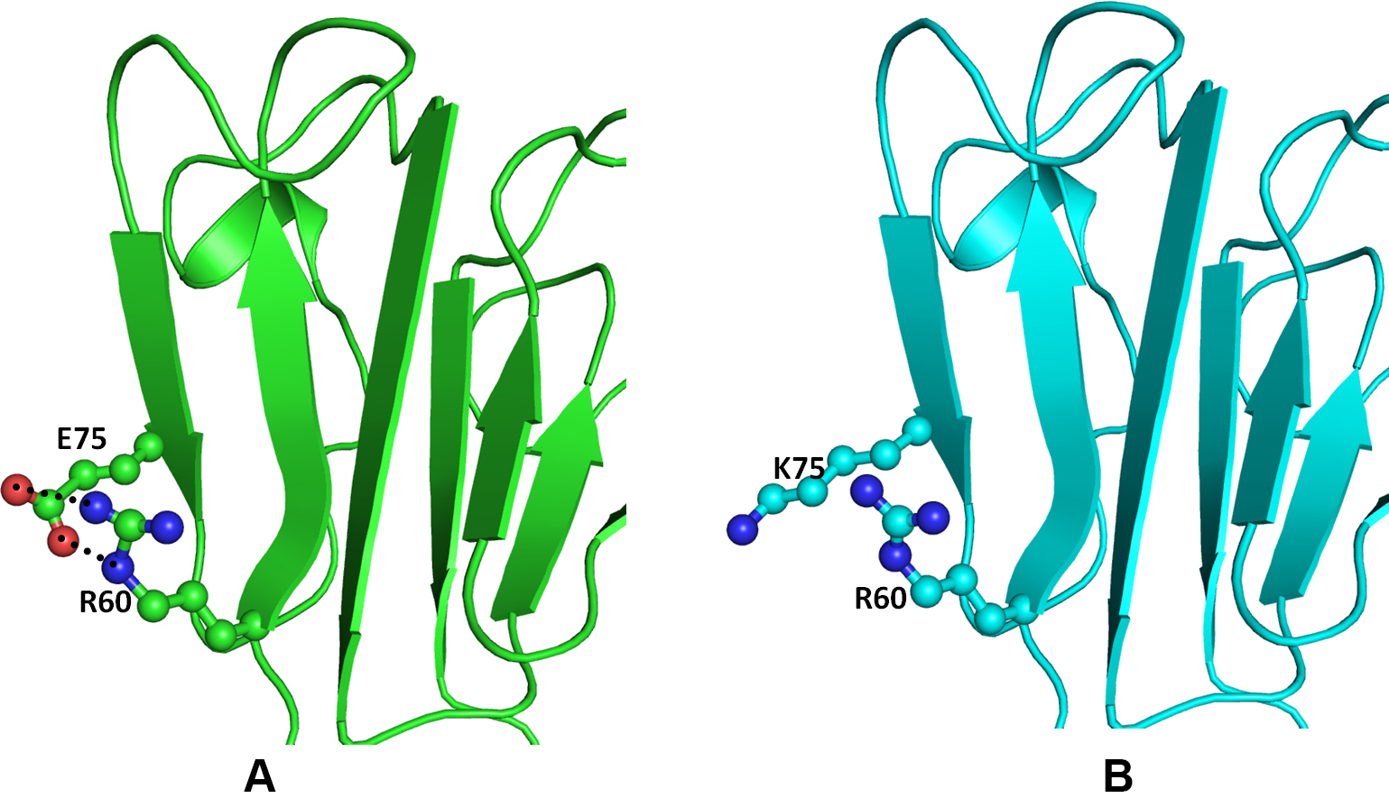
 Figure 6 of
Kumar, Mol Vis 2013; 19:2436-2450.
Figure 6 of
Kumar, Mol Vis 2013; 19:2436-2450.