Figure 5 of
Kumar, Mol Vis 2013; 19:2436-2450.
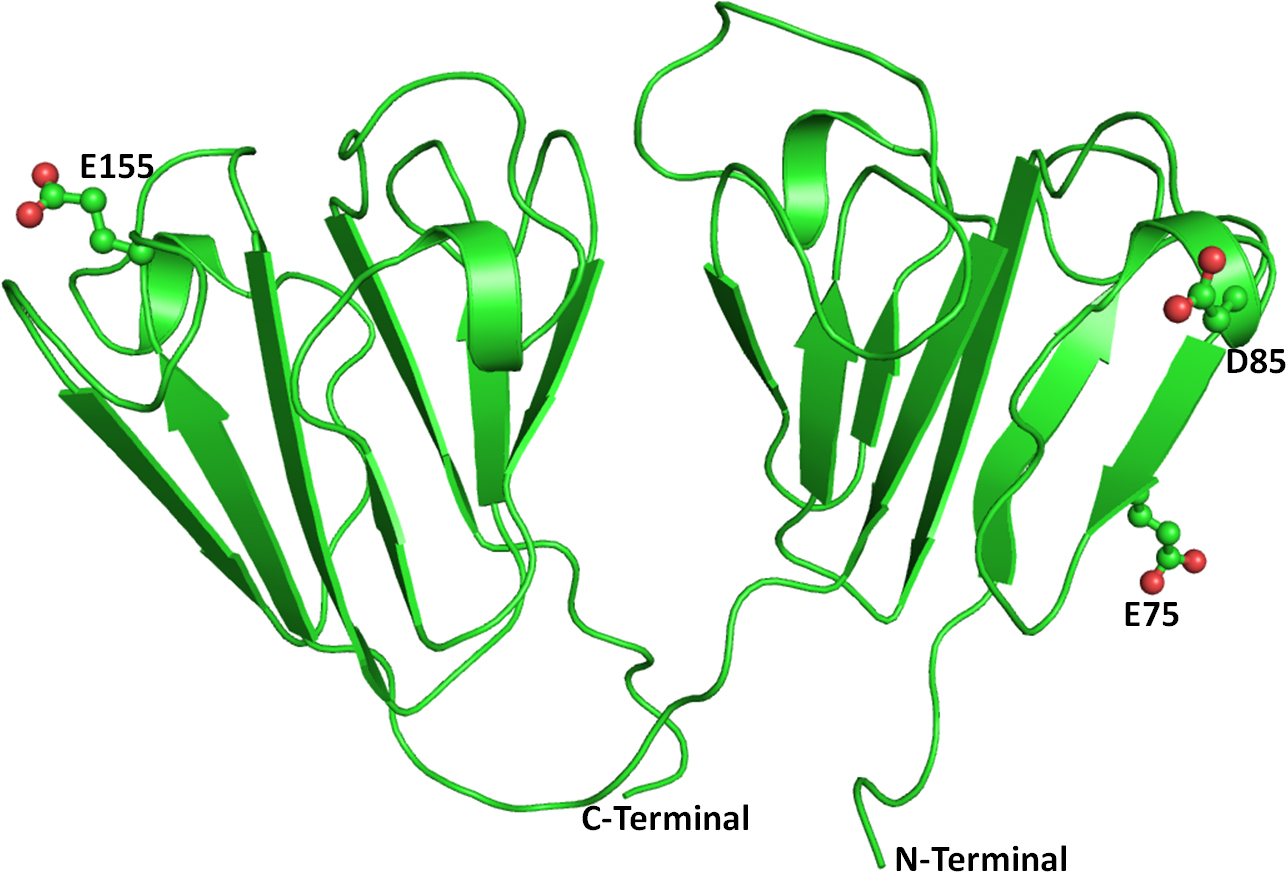
Figure 5.
Cartoon representation of the crystal structure of the wild-type beta crystallin B1 protein. The residues at the mutation site are shown as balls and sticks.
 Figure 5 of
Kumar, Mol Vis 2013; 19:2436-2450.
Figure 5 of
Kumar, Mol Vis 2013; 19:2436-2450.