Figure 2. Cartoon representation of the model structure of the wild-type crystallin beta-A4 protein. The disulfide bridge and residues
at the mutation site are shown as balls and sticks. The newly generated loops (residue 83–87 and 180–183) are in magenta.
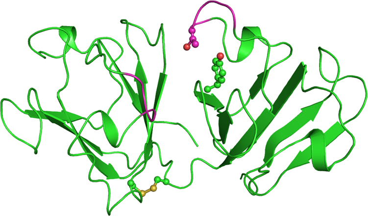
 Figure 2 of
Kumar, Mol Vis 2013; 19:2436-2450.
Figure 2 of
Kumar, Mol Vis 2013; 19:2436-2450.