Figure 4. Electron microscopy antibody-labeling of platelet-derived growth factor receptor alpha in a normal human cornea. Strong labeling
was seen in all layers of the epithelium (A). Labeling was also evident in the stromal keratocytes, especially their cell membranes (B) and in the surrounding extracellular matrix (C). Descemet’s membrane (D) and the endothelial monolayer (E) exhibited weaker labeling. No labeling was observed in control sections in which the primary antibody was omitted (F). Abbreviations: DM=Descemet’s membrane, E=Epithelium, EN=Endothelium, KR=Keratocyte, S=Stroma.
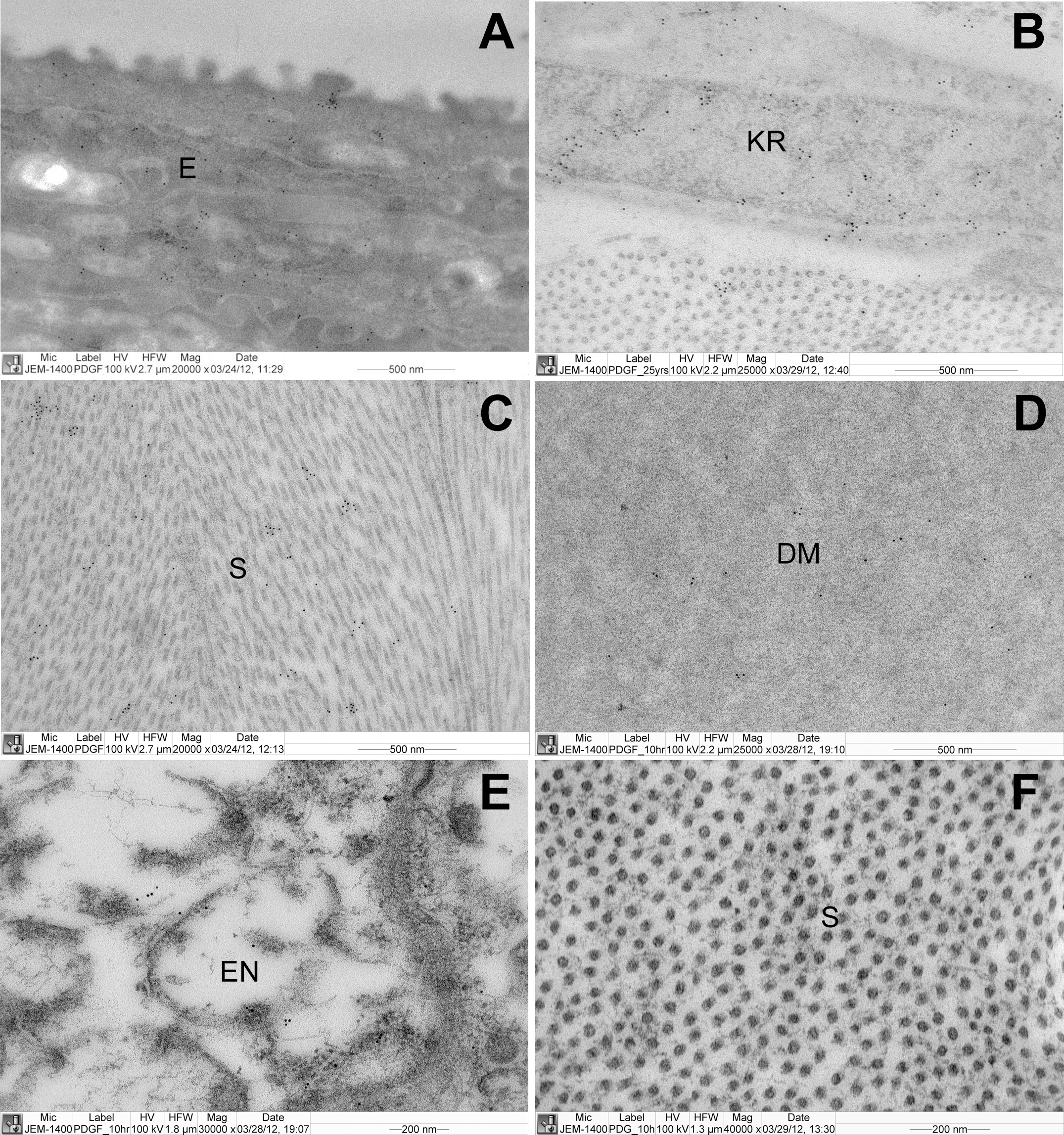
 Figure 4 of
Guggenheim, Mol Vis 2013; 19:243-253.
Figure 4 of
Guggenheim, Mol Vis 2013; 19:243-253.