Figure 2. Ophthalmological findings in patients from the two families. A: The fundus appearance of the right eye of patient III:3 in family RP24 shows atrophic retinal pigment epithelial changes,
attenuation of the retinal vessels, and irregular pigment clumps in the peripheral retina. B: A slit-lamp photograph of the same eye showed dense white opacities located in the central zone of the lens. C: The fundus appearance of the right eye of patient III:1 in family RP106 shows atrophic retinal pigment epithelial changes,
attenuation of the retinal vessels, and irregular pigment clumps in the peripheral retina. D: A photograph of the anterior segment of the same eye shows the opacities in the central zone of the lens. E: Fundus appearance of the right eye of individual III:6 from family RP106 displays a normal appearance. F: Electroretinography (ERG) shows normal rod and cone cell responses. G: The fundus appearance of the right eye of patient IV:3 (daughter of individual III:6) presents atrophic retinal pigment
epithelial changes. H: The ERG shows extinguished rod and cone cell responses.
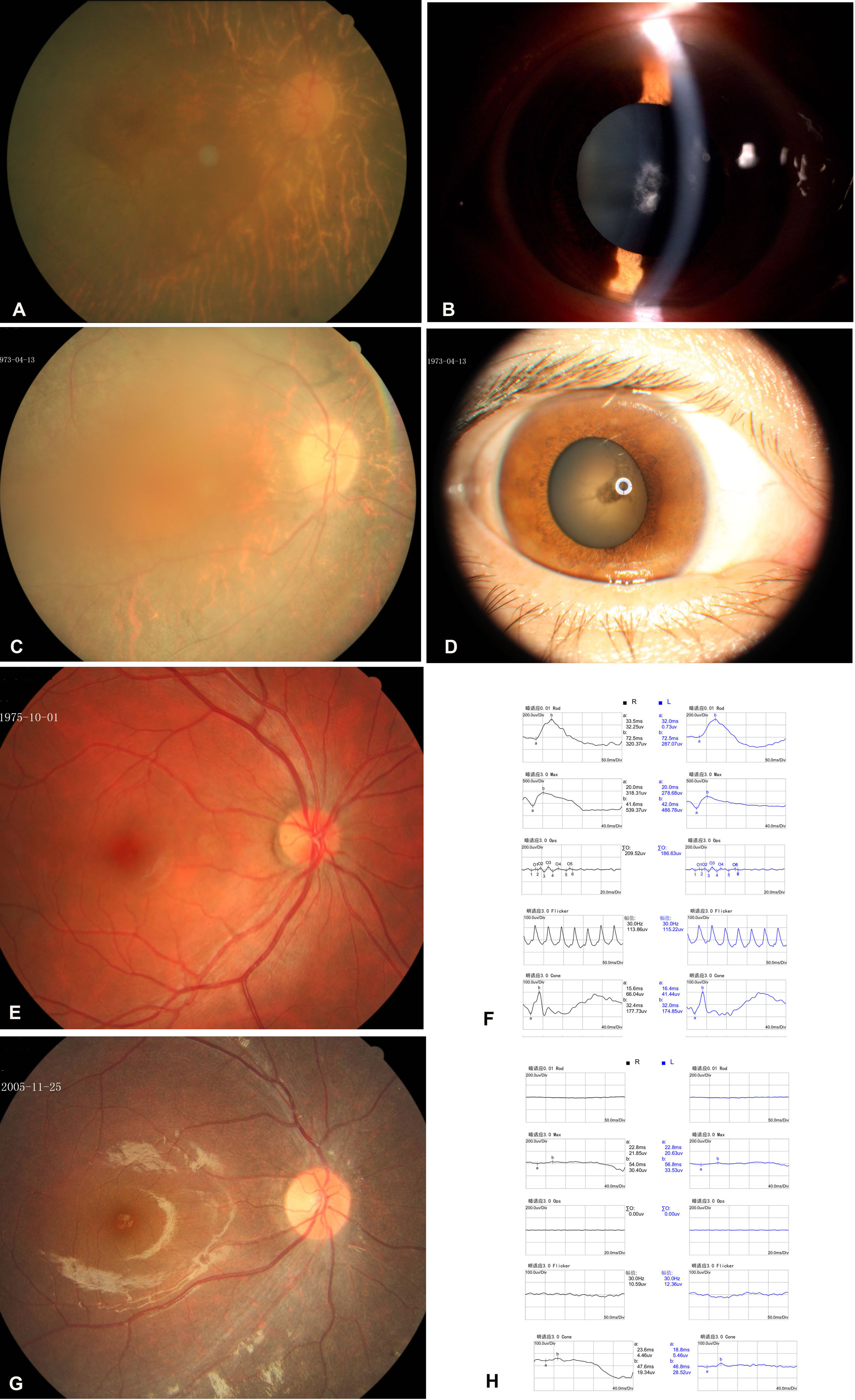
 Figure 2 of
Dong, Mol Vis 2013; 19:2426-2435.
Figure 2 of
Dong, Mol Vis 2013; 19:2426-2435.