Figure 3. Fundus photographs from individuals with small nuclear riboprotein 200 kDa mutations. A: UTAD565-02 at age 59 with moderatively advanced retinitis pigmentosa . Her right eye fundus shows diffuse atrophy of the
optic nerve and the retina outside the macula with heavy pigmentary deposits in the equator and vascular attenuation, while
the macula area shows intact retinal pigment epithelium with no foveal reflex. B: RFS048–4884 at age 40 showed severe vascular attenuation and disc pallor. Extensive pigmentary disturbances were seen throughout
the peripheral retina. C: RFS048-5420 at age 14 showed moderate vascular attenuation and disc pallor. Moderate pigment clumping was seen in the periphery.
D: UTAD701-01 at age 34 shows vascular attenuation, mild disc pallor, and heterogeneous, mottled fundus pigment along the temporal
arcades with preserved pigmentation in the central macula.
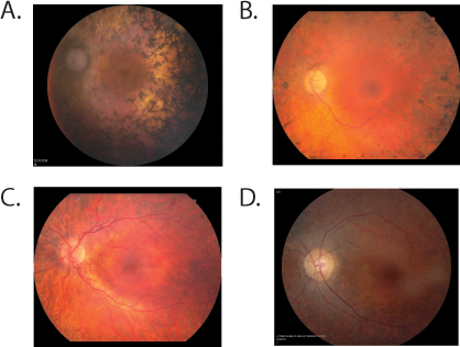
 Figure 3 of
Bowne, Mol Vis 2013; 19:2407-2417.
Figure 3 of
Bowne, Mol Vis 2013; 19:2407-2417.