Figure 5. Optic coherence tomography findings. A and B: Time domain optic coherence tomography (OCT; retinal mapping) of patient II-1 at the age of 22 years (A) and II-2 at the age of 16 years (B) show total macular thinning in both eyes. C: Spectral-domain OCT (HD-5-line raster) of patient II-2 at the age of 23 years, showing marked macular thinning with indistinguishable
retinal layers in the macular areas of both eyes.
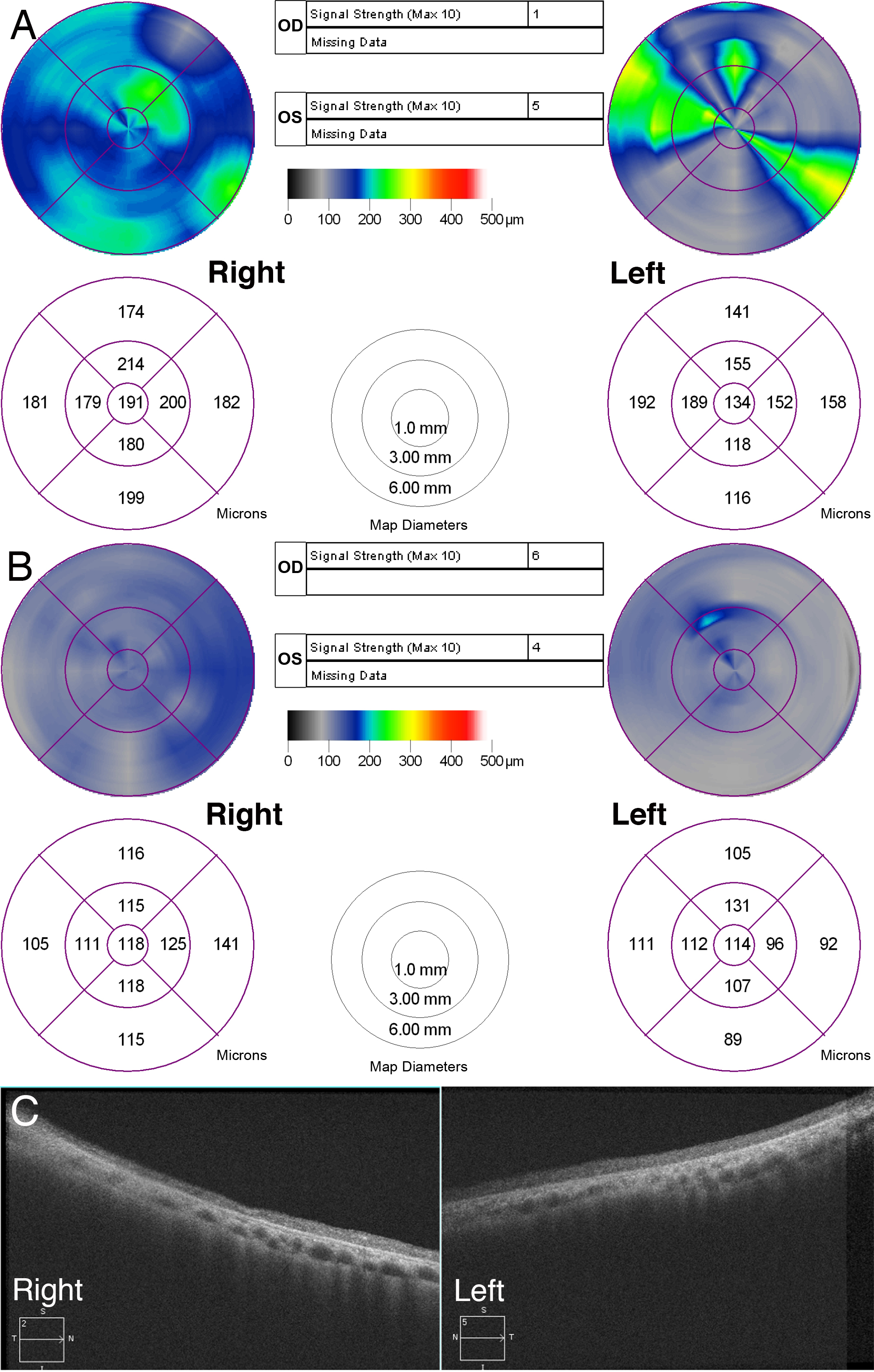
 Figure 5 of
Katagiri, Mol Vis 2013; 19:2393-2406.
Figure 5 of
Katagiri, Mol Vis 2013; 19:2393-2406.