Figure 4. Fundus photographs of patients II-1 and II-2. A and B: Fundus photographs of patient II-1 at the age of 14 years (A) and patient II-2 at the age of 8 years (B) show retinal degeneration with attenuated vessels in the posterior poles of both eyes.
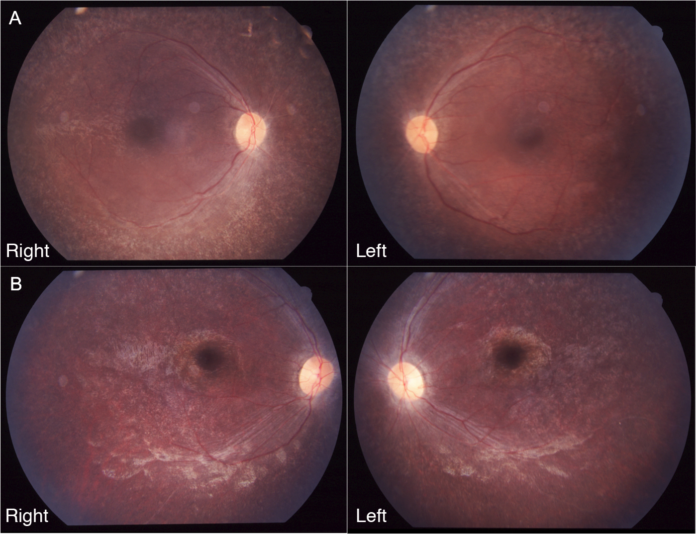
 Figure 4 of
Katagiri, Mol Vis 2013; 19:2393-2406.
Figure 4 of
Katagiri, Mol Vis 2013; 19:2393-2406.