Figure 6. Effect of EDTA and heparin on cell adhesion of retinal Müller cells. A: EDTA (EDTA) or EDTA (5 mM) or heparin (10 μg/ml) was preincubated with human retinal Müller cell line (MIO-M1) cell suspension
for 2 h and then added to the vitreous explants. Thereafter, stereomicroscopic images of MIO-M1 cells on the vitreous surface
were taken after hematoxylin and eosin (H&E) staining. The micrograph depicts cell morphology on the vitreous surface in the
magnified image. Scale bar=200 μm. B: 3-(4,5-dimethylthiazol-2-yl)-2,5-diphenyltetrazolium bromide (MTT) assay was performed to evaluate the number of adherent
cells on the vitreous surface. Data are expressed as mean±standard deviation (SD; n=4). *, significantly different from the
cell-treated group (p<0.05).
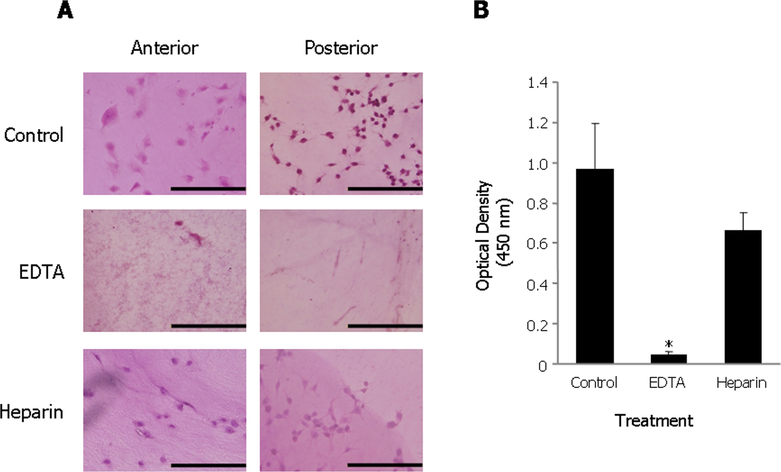
 Figure 6 of
Oki, Mol Vis 2013; 19:2374-2384.
Figure 6 of
Oki, Mol Vis 2013; 19:2374-2384.