Figure 4. Morphologies of retinal Müller cells contracting vitreous explants from chicken embryos. Stereomicroscopic images of human
retinal Müller cell line (MIO-M1) cells on the vitreous surface were taken after hematoxylin and eosin (H&E) staining at the
indicated periods. The micrograph depicts cell morphology on the vitreous surface at the anterior (A) or the posterior region (B) and the magnified image is shown in (C) or (D), respectively. The magnified image (E or F) is the cell morphology on the vitreous surface at the anterior or the posterior region, respectively, at 6 h after cell
adhesion. Scale bar=200 μm.
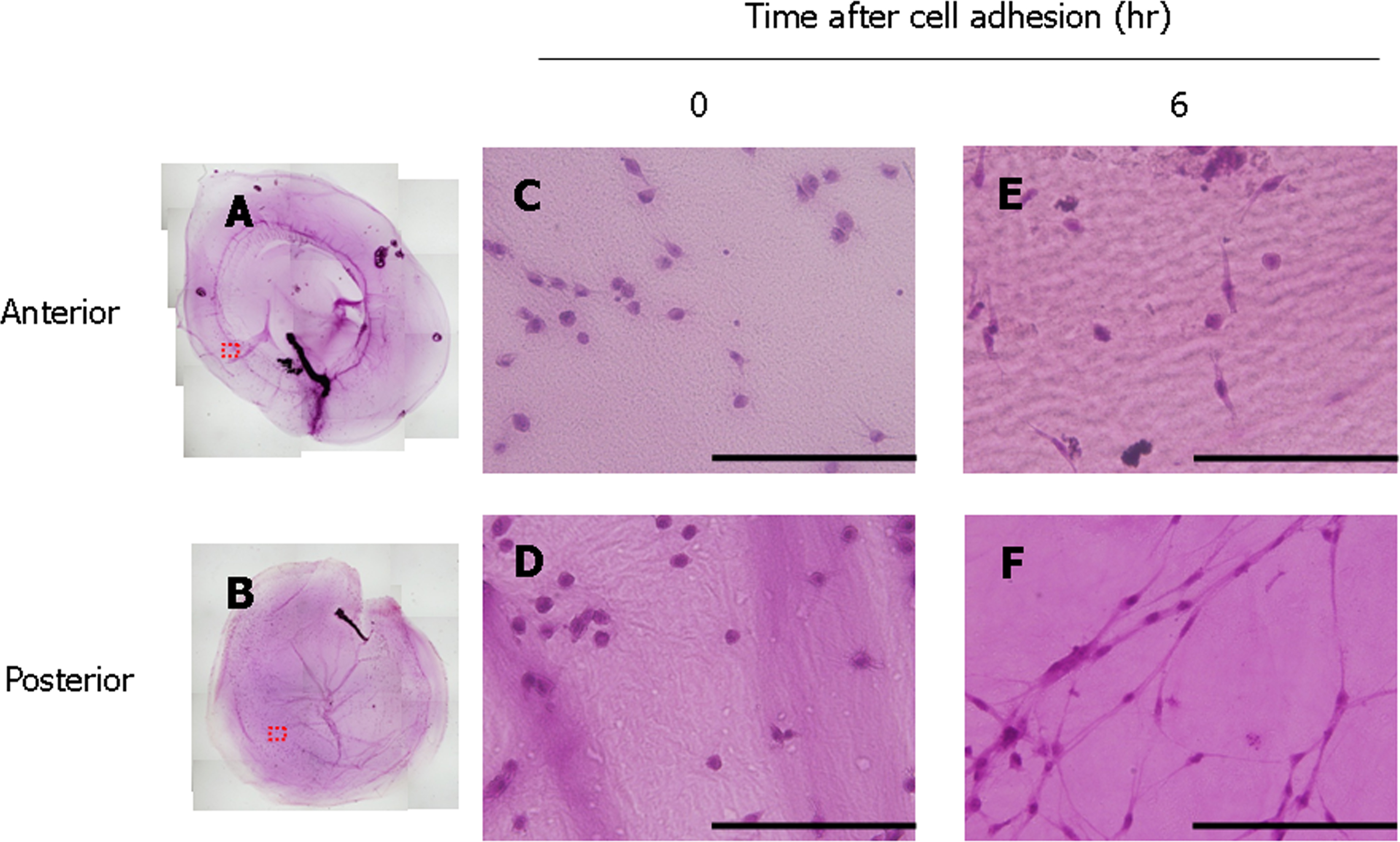
 Figure 4 of
Oki, Mol Vis 2013; 19:2374-2384.
Figure 4 of
Oki, Mol Vis 2013; 19:2374-2384.