Figure 3. Vitreous contraction analysis of endothelial cells in vitreous explants from chicken embryos. A: After cell adhesion of bEnd.3 cells, the vitreous explants were incubated in Dulbecco’s modified Eagle’s medium (DMEM),
in the presence or absence of fetal bovine serum (FBS), at 37 °C up to the indicated periods. Thereafter, the wet weight of
vitreous explants was measured for the quantitative analysis of vitreous contraction. Control vitreous explants were incubated
at 37 °C in the culture medium for 24 h in the absence of cells. B: Vitreous explants were cultured with adherent bEnd.3 cells under the same conditions. Thereafter, the vitreous explants
were stained with Coomassie brilliant blue (CBB) to evaluate the macroscopic effects of cell adhesion. Data are expressed
as mean±standard deviation (SD; n=4). *, significantly different from cell-free control group (p<0.05). Not significant versus
cell without FBS.
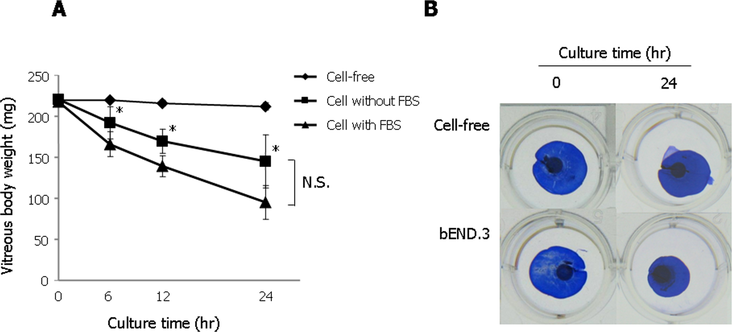
 Figure 3 of
Oki, Mol Vis 2013; 19:2374-2384.
Figure 3 of
Oki, Mol Vis 2013; 19:2374-2384.