Figure 2. Vitreous contraction analysis of retinal Müller cells in vitreous explants from chicken embryos. A: After cell adhesion of human retinal Müller cell line (MIO-M1) cells, the vitreous explants were incubated in Dulbecco’s
modified Eagle’s medium (DMEM), in the presence or absence of fetal bovine serum (FBS), at 37 °C up to the indicated periods.
The wet weight of vitreous explants was measured for the quantitative analysis of vitreous contraction. Control vitreous explants
were incubated at 37 °C in the culture medium for 24 h in the absence of cells. B: Vitreous explants were cultured with adherent MIO-M1 cells under the same conditions. Thereafter, the vitreous explants
were stained with Coomassie brilliant blue (CBB) to evaluate the macroscopic effects of cell adhesion. Data are expressed
as mean±standard deviation (SD; n=4). *, significantly different from cell-free control group (p<0.05). Not significant versus
cell without FBS.
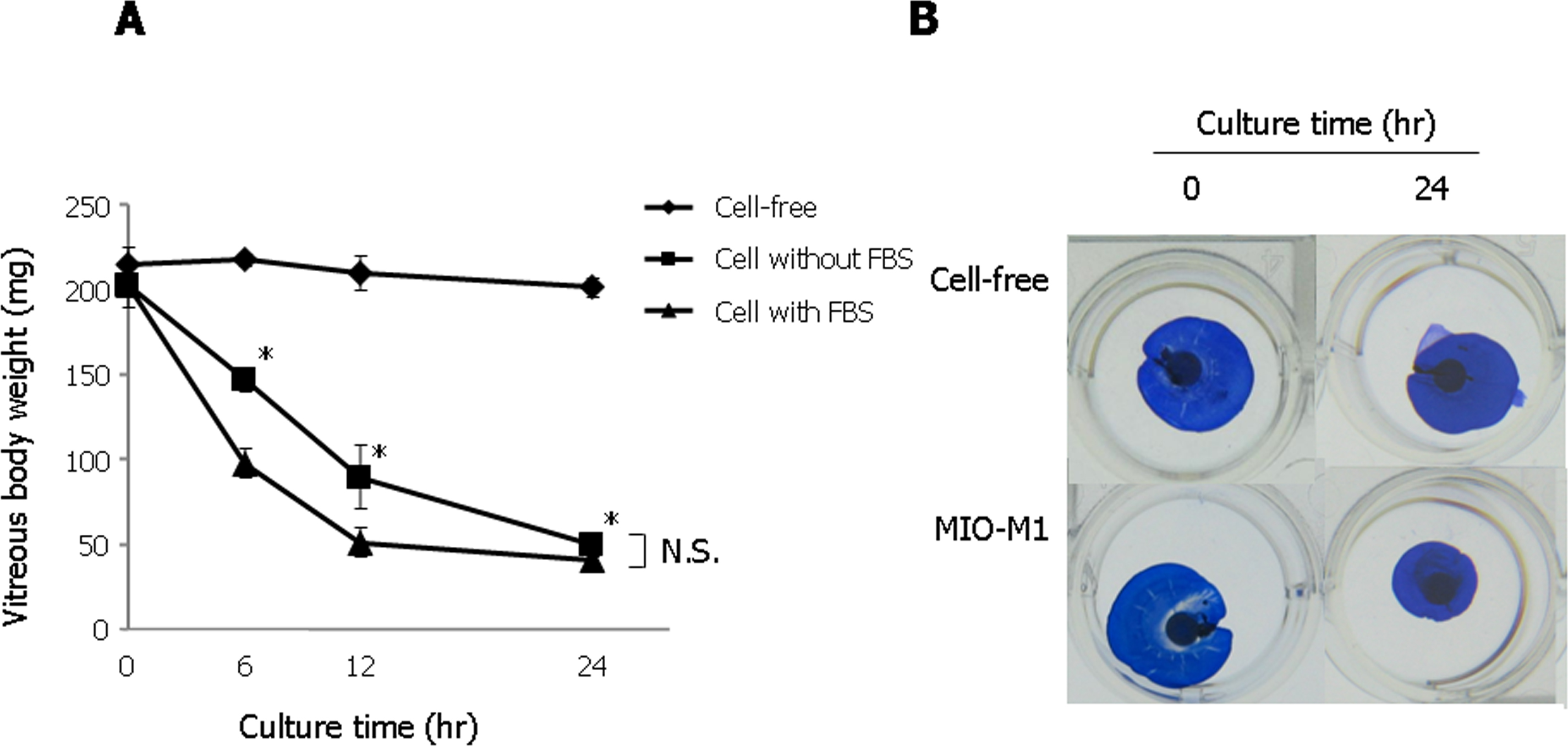
 Figure 2 of
Oki, Mol Vis 2013; 19:2374-2384.
Figure 2 of
Oki, Mol Vis 2013; 19:2374-2384.