Figure 5. Intraocular pressure in wild-type and hypoxia-inducible factor-1α retinal ganglion cell-knockout mice with and without repetitive
hypoxic preconditioning. Intraocular pressure (IOP) in the experimental eyes (filled symbols) and respective fellow eyes (open
symbols) in the wild-type (WT; red; n=10) and hypoxia-inducible factor-1α retinal ganglion cell-knockout (HIF-1α RGC-KO; purple; n=8) groups without repetitive hypoxic preconditioning (RHP), and the WT (green; n=8) and HIF-1α RGC-KO (blue; n=14) with prior RHP over the 3-week period of intraocular hypertension are shown. All IOP values for the experimental
eyes in each group at 24 h and 1, 2, and 3 weeks were significantly different from their respective baselines and from their
fellow eyes at each time point, but no statistically significant differences were noted among the four groups. Mean±standard
error of the mean (SEM).
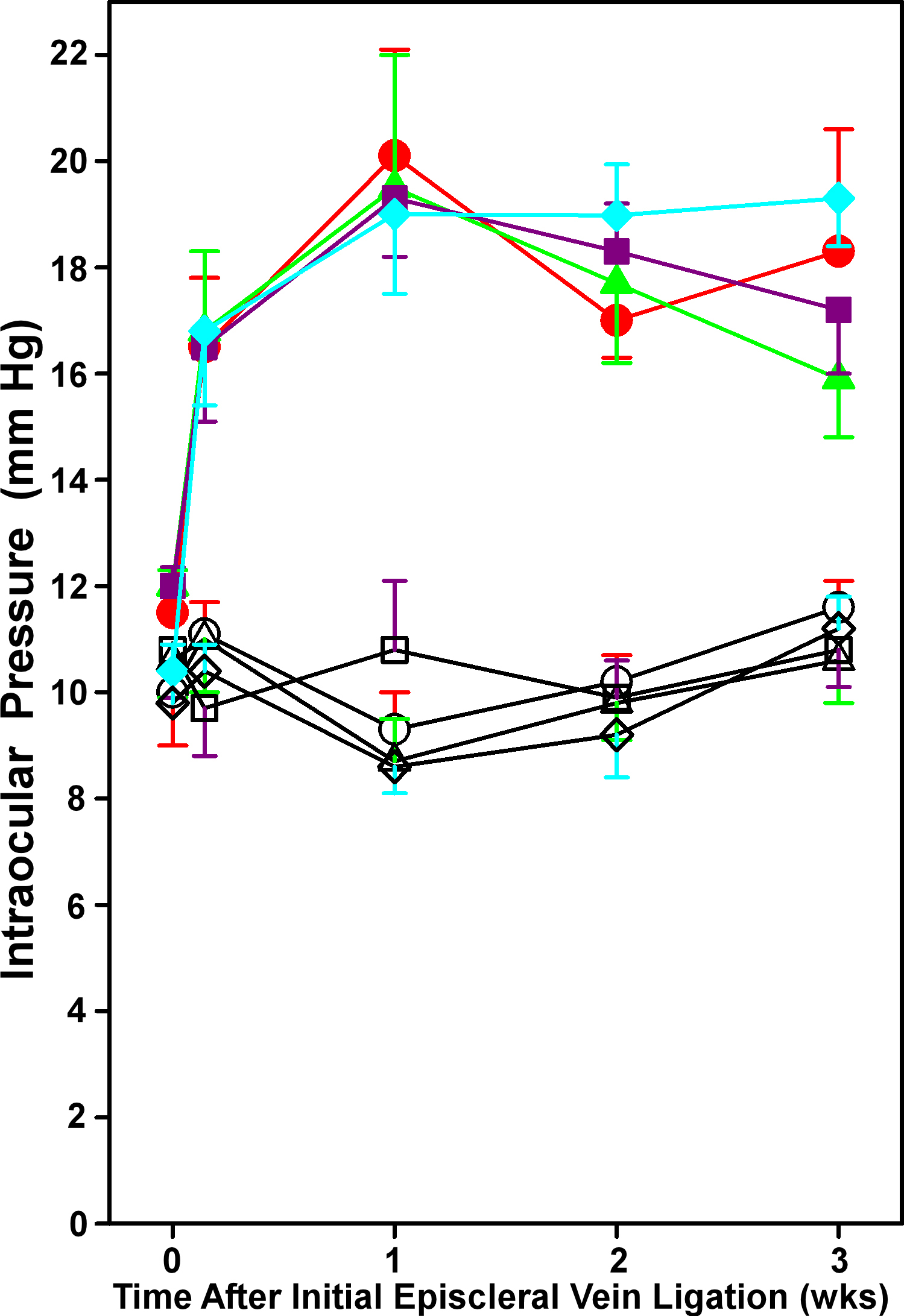
 Figure 5 of
Zhu, Mol Vis 2013; 19:2360-2372.
Figure 5 of
Zhu, Mol Vis 2013; 19:2360-2372.