Figure 6. Structural details of
cortical fiber cells. A: Displayed is a low
magnification overview of the capsule, epithelium (N, nuclei),
and outer cortex from 2011-406 (92/M). B: A high
magnification image shows an epithelial cell nuclear envelope
with a nuclear pore (arrow) and endoplasmic reticulum with
adherent ribosomes (arrowheads) from 2011- 523 (22/M). C:
High magnification image of a mitochondrion with a cristae
membrane labeled (arrow) from 2011-523 (22/M). D: An
intermediate magnification image shows elongating fiber cells
and regular fiber cells of the outer cortex, just posterior to
the equatorial plane. A gap junction between elongating fiber
cells is marked (arrowhead). A 10-µm-thick intact and
undistorted capsule is shown (double arrow) from 2011-565
(55/M). E: A well-preserved interface is shown between
Cell 1, a dark staining elongating fiber cell, and Cell 2
showing a gap junction (paired membranes) from 2011-565 (55/M).
F: A similar gap junction (paired membranes) between two
fiber cells (cells 1 and 2) is shown deeper within the outer
cortex from 2011-565 (55/M).
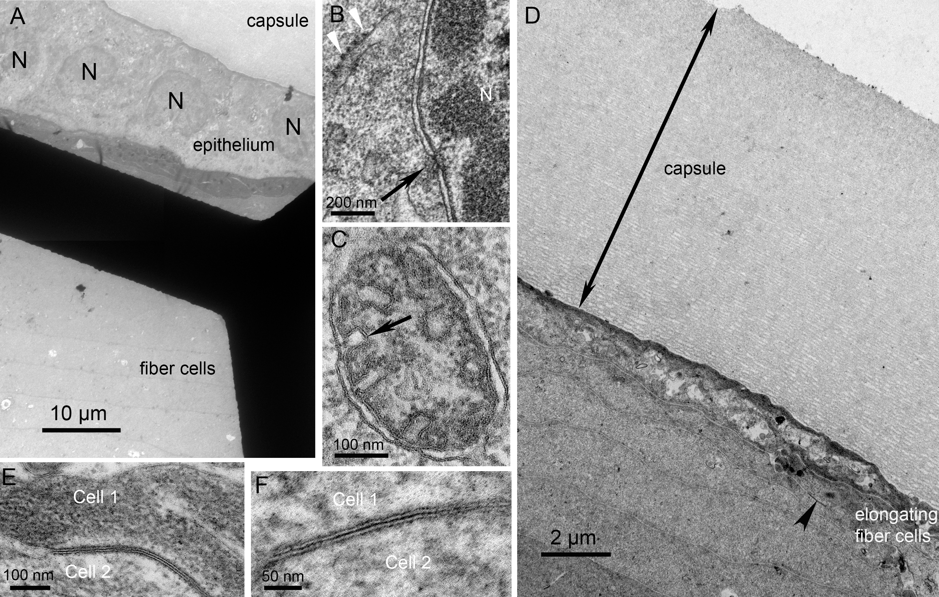
 Figure 6
of Mohamed, Mol Vis 2013; 19:2352-2359.
Figure 6
of Mohamed, Mol Vis 2013; 19:2352-2359.