Figure 5. Electron micrographs of multilamellar bodies display their essential features. A: A multilamellar body 1.4 μm in diameter was observed in a donor lens: 2011–406 (92/M). Note the absence of membrane layers
and the large electron lucent rim indicating a mature particle. B: A multilamellar body 1.7 μm in diameter observed in a cataractous nucleus: 4–6-11#2 (69/M). Note the prominent multilamellar
membranes, which are partially disrupted (arrowheads) indicating an intermediate stage of maturation. The repeat period of
the bilayer membranes is about 5 nm for the four bilayers shown at high magnification (inset).
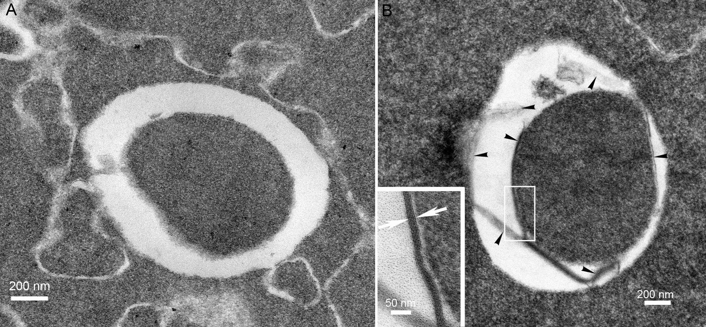
 Figure 5 of
Mohamed, Mol Vis 2013; 19:2352-2359.
Figure 5 of
Mohamed, Mol Vis 2013; 19:2352-2359.