Figure 3. Low magnification electron micrographs of fetal and embryonic nuclear fiber cells. A: Donor lens image is from the fetal nucleus close to the embryonic nucleus: 2011–406 (92/M). B: Cataract nucleus image is from an opaque region of the embryonic nucleus: 3–25–11#3 (74/F). The open space between cells
2 and 3 is typically the location of a gap junction (GJ, arrow). [Key, here and subsequent figures: Sample ID (age in years/gender)].
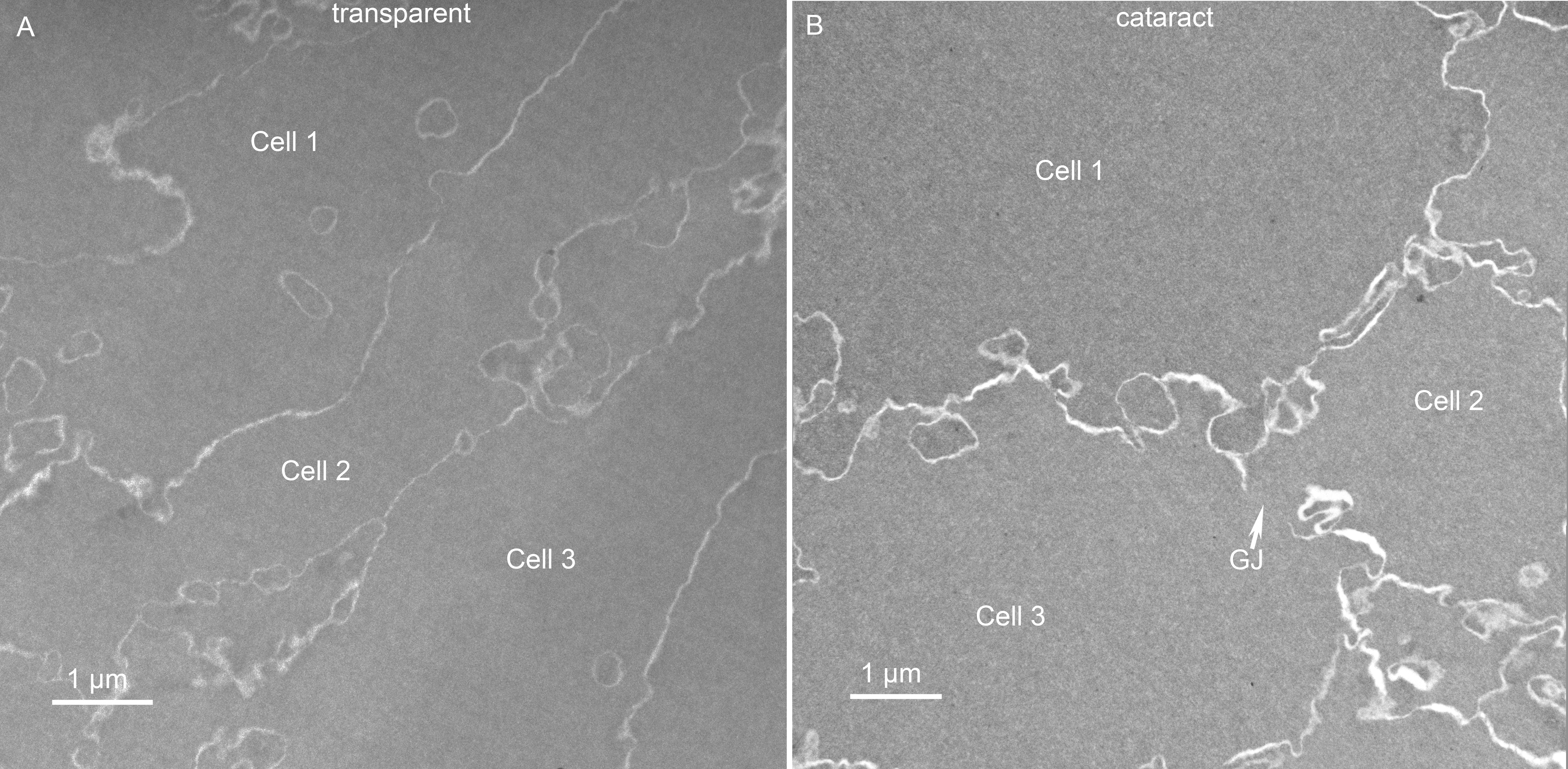
 Figure 3 of
Mohamed, Mol Vis 2013; 19:2352-2359.
Figure 3 of
Mohamed, Mol Vis 2013; 19:2352-2359.