Figure 3. The morphological changes observed in retinal pigment epithelium (RPE) cultures treated with human amniotic fluid (HAF) for
over 30 days. A: HAF-treated RPE culture after 3 days. The cells grew as a monolayer and were arranged in parallel to each other. B: After 7 days, the cells began to form cell clusters. C, D: The RPE-derived neurosphere-like colonies formed and increased in number during the 2nd and 3rd weeks. E: Consequently, the RPE-derived colonies detached from the base of the plate and floated into the medium. F, G, H, I, J: The control cultures (RPE cells cultured in Dulbecco's modified Eagle medium: Nutrient mixture F-12 [DMEM/F12]) had a considerable
delay in forming neurosphere-like colonies.
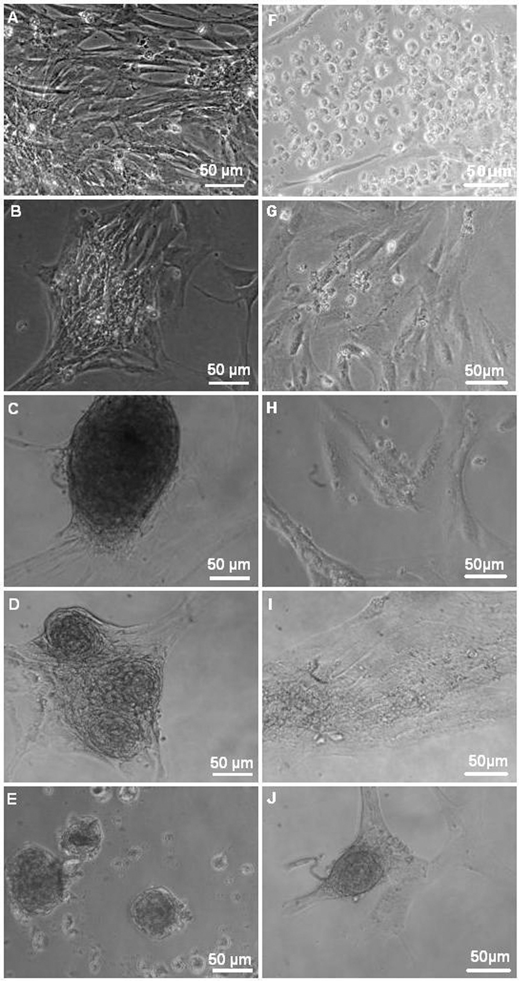
 Figure 3 of
Davari, Mol Vis 2013; 19:2330-2342.
Figure 3 of
Davari, Mol Vis 2013; 19:2330-2342.