Figure 1. The different stages of retinal pigment epithelium (RPE) cell growth in culture. A, B: RPE cells freshly isolated from a human eye. C: Up to passage number 5, RPE cells have an elongated morphology containing several long processes. D: Confluent RPE cells in culture organized into cobblestone structures. E, F: RPE-derived colonies under normal culture conditions. G, H: The appearance of flattened cells in an aged culture (over passage 5) suggests that senescence is occurring.
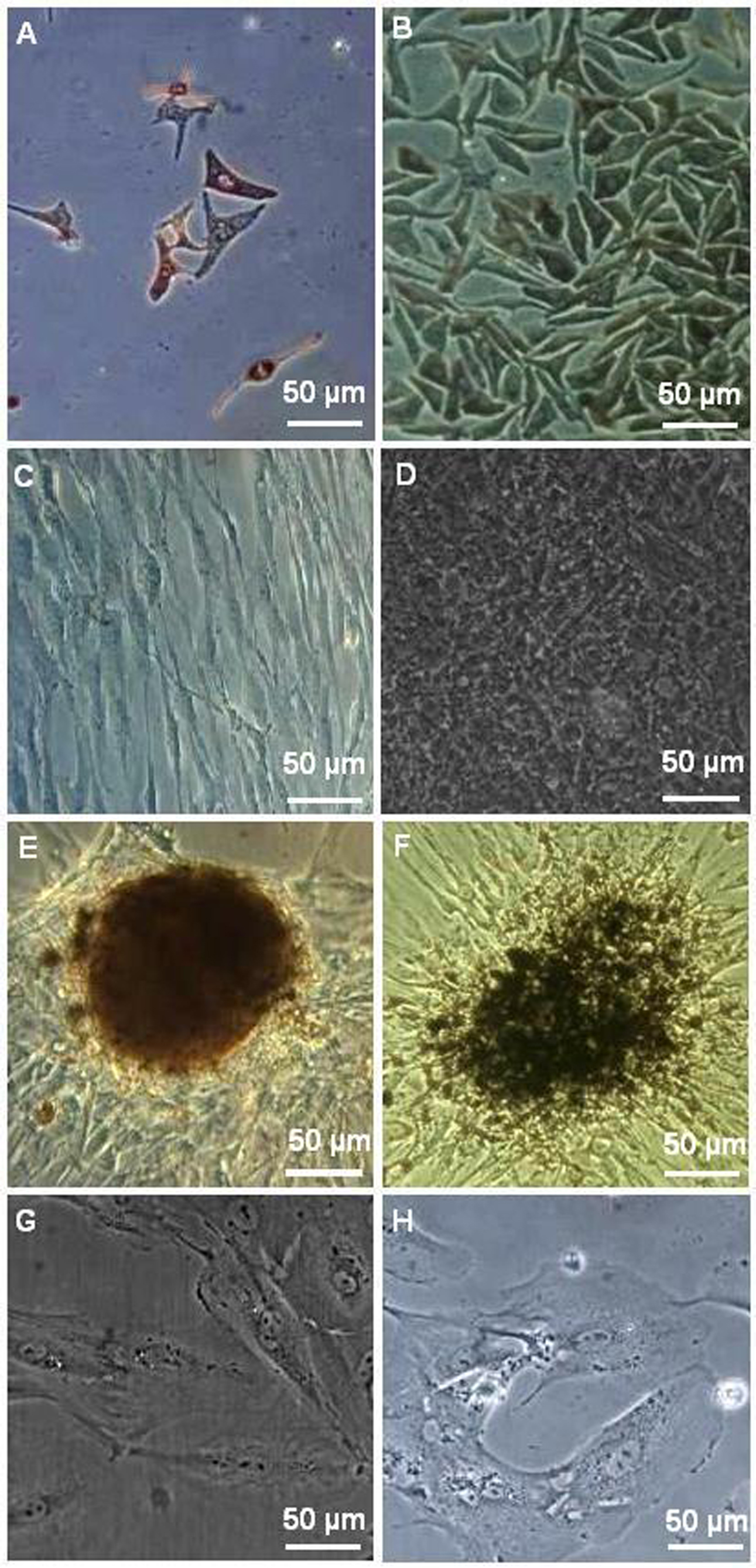
 Figure 1 of
Davari, Mol Vis 2013; 19:2330-2342.
Figure 1 of
Davari, Mol Vis 2013; 19:2330-2342.