Figure 5. Stra6 protein expression in the P14 mouse retina. Immunohistochemical analysis was performed with a rabbit polyclonal serum
raised against the C-terminus of Stra6 (red), on retinal sections of wild-type (A), Rpe65−/− (B), Cspg5−/−, and (C) Cspg5−/−/Rpe65−/− (D) mice. Stra6 was predominantly expressed at the basolateral membrane of the retinal pigment epithelium (RPE) in all analyzed
genotypes. The nuclei were stained in blue with 4',6-diamidino-2-phenylindole dihydrochloride (DAPI), and the images were
merged. As a negative control, a serum of a non-immunized rabbit was used, the nuclei stained with DAPI, and the images merged
(E). Abbreviations: retinal pigment epithelium (rpe); photoreceptor outer segments (os); photoreceptor inner segments (is);
outer nuclear layer (onl). Scale bar equals 40 μm.
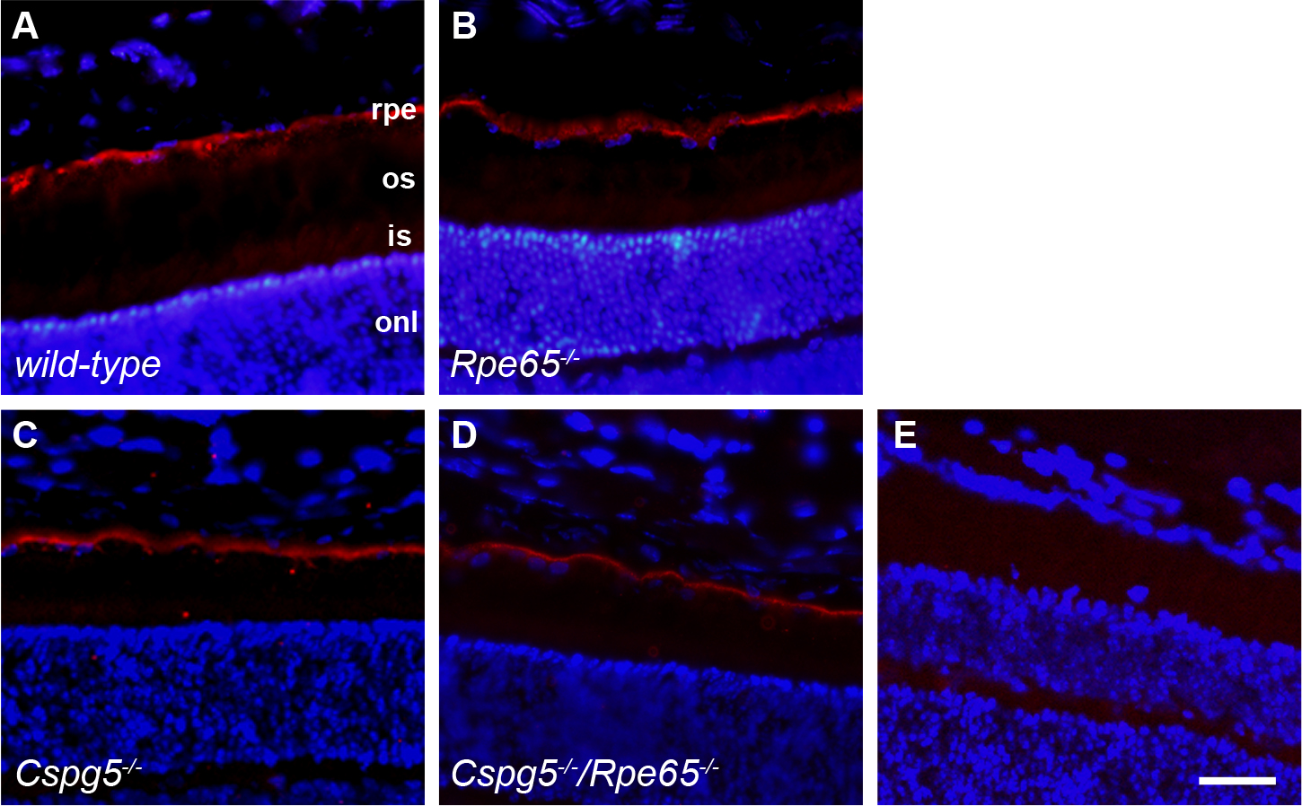
 Figure 5 of
Cottet, Mol Vis 2013; 19:2312-2320.
Figure 5 of
Cottet, Mol Vis 2013; 19:2312-2320.