Figure 2. Cone degeneration in Rpe65−/− and Cspg5−/−/Rpe65−/− retinas. Central regions of retinas from 8-week-old wild-type (A), Cspg5−/− (B), Rpe65−/−, and (C) Cspg5−/−/Rpe65−/− (D) mice were analyzed. Cones were labeled with fluorescein-conjugated peanut agglutinin (green), nuclei were stained with 4',6-diamidino-2-phenylindole
dihydrochloride (blue), and the images were merged. The cone outer segments were prominently stained in the wild-type and
Cspg5−/− retinas (A, B). Residual staining of cone inner segments was observed in the central retina of the Rpe65−/− and Cspg5−/−/Rpe65−/− mice (C, D). In the peripheral regions of the Cspg5−/−/Rpe65−/− retinas, cone outer segment labeling was still observed, with mislocalization to the synaptic endfeet of cone photoreceptors
(star; E). Abbreviations: photoreceptor outer segments (os); photoreceptor inner segments (is); outer nuclear layer (onl); outer plexiform
layer (opl); inner nuclear layer (inl); inner plexiform layer (ipl); ganglion cell layer (gcl). Scale bar equals 50 μm.
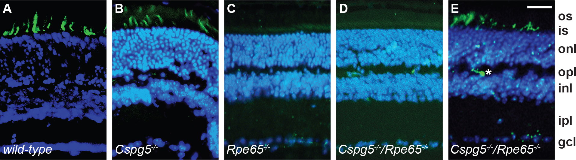
 Figure 2 of
Cottet, Mol Vis 2013; 19:2312-2320.
Figure 2 of
Cottet, Mol Vis 2013; 19:2312-2320.