Figure 1. Progression of cone degeneration in retinal pigment epithelium protein of 65 kDa (Rpe65)−/− and Cspg5−/−/Rpe65−/− retinas. Fluorescein-conjugated peanut agglutinin (FITC-PNA)-labeled cones were counted on retinal sections of 2-, 4-, and
8-week-old mice. Cones had almost disappeared in the central retina of the 4- and 8-week-old Rpe65−/− and Cspg5−/−/Rpe65−/− mice. The peripheral region considered for cone counting in the Rpe65−/− and Cspg5−/−/Rpe65−/− mice was located toward the edges of the retina. With two-way ANOVA using factors of genotype and age, no significant differences
were found between the Rpe65−/− and Cspg5−/−/Rpe65−/− mice in the number of cones, in the central and peripheral retina (n=4). For all time points, the number of cones per field
of view±standard error of the mean (SEM) is shown.
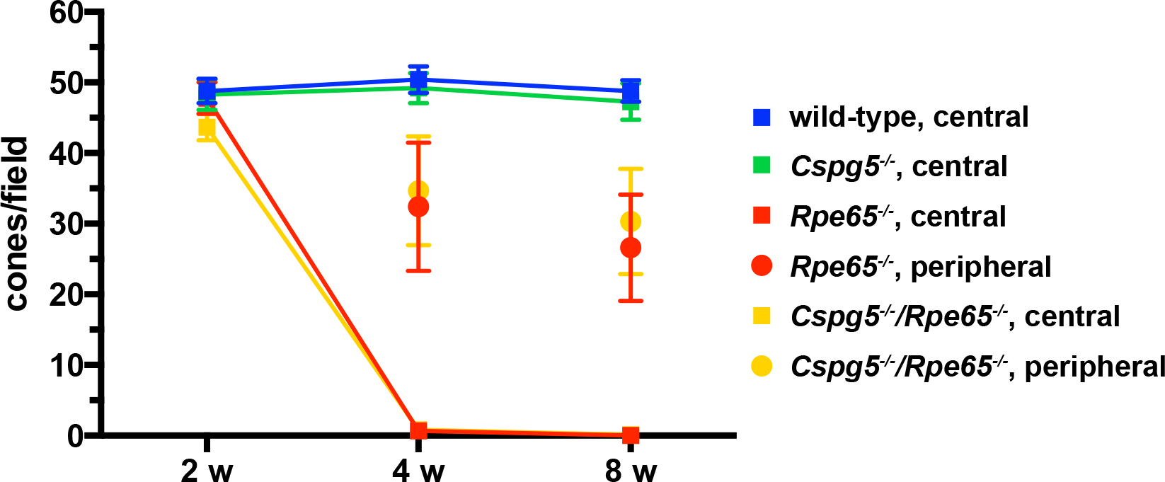
 Figure 1 of
Cottet, Mol Vis 2013; 19:2312-2320.
Figure 1 of
Cottet, Mol Vis 2013; 19:2312-2320.