Figure 3. Goldmann visual fields of both eyes of each patient using target sizes: I4e, III4e, and V4e. The left-hand column shows the
fields of the left eye, while the right-hand column illustrates the fields of the right eye. Case 1 has a relatively large
central scotoma; the left eye shows smaller paracentral islands of sensitivity loss. Case 2 has normal fields. Case 3 has
a left central and paracentral relative scotomata to static stimuli only. The right eye field shows a small relative scotoma.
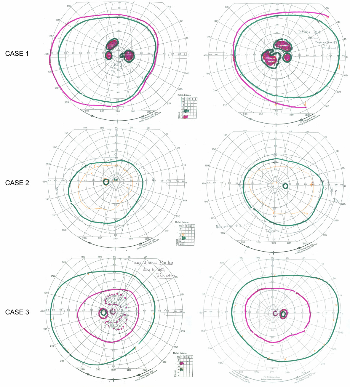
 Figure 3 of
Ba-Abbad, Mol Vis 2013; 19:2250-2259.
Figure 3 of
Ba-Abbad, Mol Vis 2013; 19:2250-2259.