Figure 2. Color fundus photographs , fundus autofluorescence images, and optical coherence tomography images of cases 1, 2, and 3. A: Case 1-A mild pigment atrophy in the central macula and hyperpigmentation nasal to the fovea. B: Speckled hypoautofluorescence in the macular region. C: Irregular reflectivity at the level of the outer retinal layers.
D: Case 2-Subtle pigment changes at the center of the macula. E: Normal fundus autofluorescence (FAF). F: Case 3-The retina appears normal except for a central area of dense hyperpigmentation. G: A ring of hyperautofluorescence surrounding a central bull’s eye lesion. H: An elevated lesion is present at the center of the fovea. There is distortion of the retinal layers over and adjacent to
the lesion.
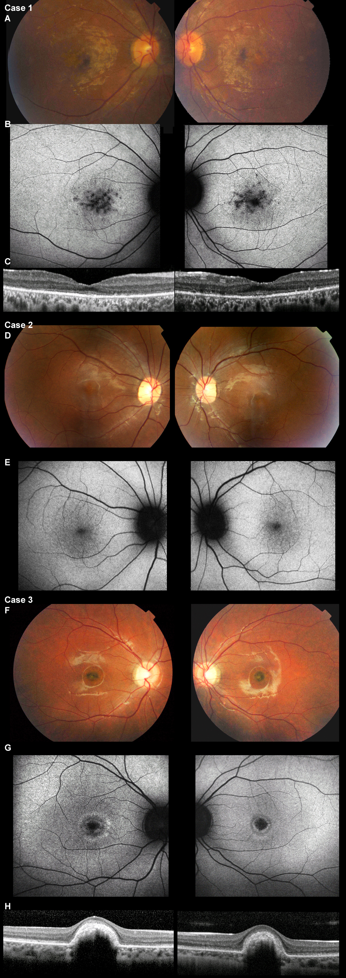
 Figure 2 of
Ba-Abbad, Mol Vis 2013; 19:2250-2259.
Figure 2 of
Ba-Abbad, Mol Vis 2013; 19:2250-2259.