Figure 2. Apelin-induced phosphorylation of Erk and Akt. A, B, C: RPE cells were incubated with 10−8 M, 10−7 M, and 10−6 M apelin for 30 min for assay of Erk and Akt phosphorylation, levels of phosphorylated and total Akt and Erk were determined
with western blot analysis, respectively. RPE cells were pretreated with 10 μM LY294002 (D, E) or 20 μM PD98059 (F, G) for 30 min and then incubated with 10−7 M apelin for 30 min for assay of Akt and Erk phosphorylation. Levels of phosphorylated Akt and Erk were determined with western
blot analysis. The data represent the mean±standard deviation (SD) of three independent experiments, *p<0.001 versus untreated
control.
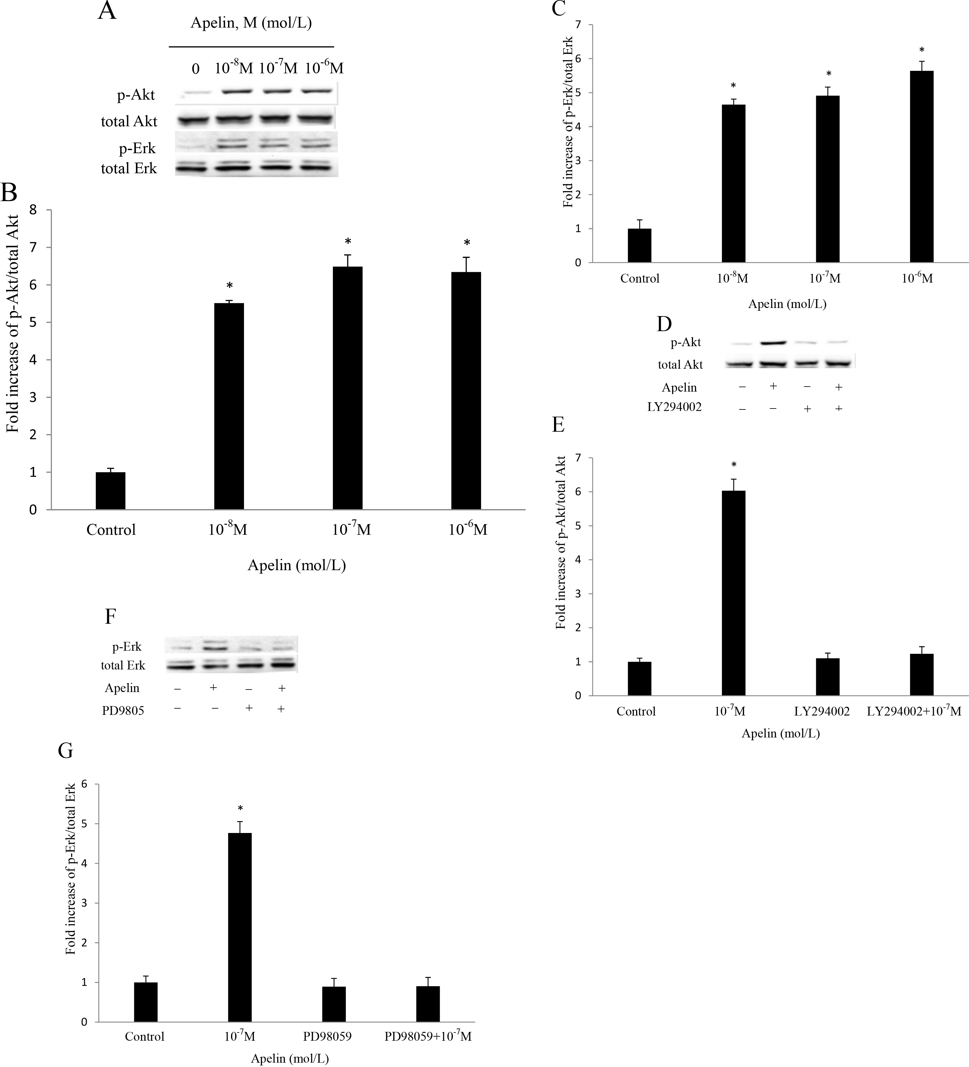
 Figure 2 of
Qin, Mol Vis 2013; 19:2227-2236.
Figure 2 of
Qin, Mol Vis 2013; 19:2227-2236.