Figure 1. Fundus photographs and optical coherence tomography scans demonstrated foveal schisis. A: Fundal exam (OD) in patient 645. B: Fundal exam (OS) in patient 645. C: Fundal exam (OD) in patient 22. D: Fundal exam (OS) in patient 22. E, F: Optical coherence tomograms through a horizontal section of the left and right eyes of patient 645 showed classic foveal
schisis.
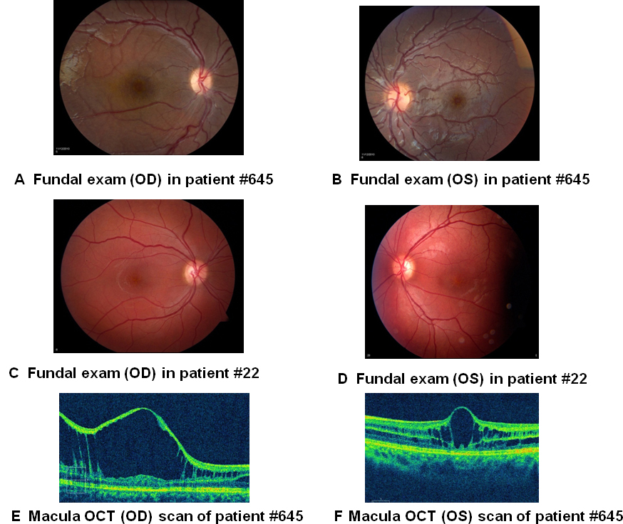
 Figure 1 of
D’Souza, Mol Vis 2013; 19:2209-2216.
Figure 1 of
D’Souza, Mol Vis 2013; 19:2209-2216.