Figure 5. Representative photomicrographs of double immunostaining of CD68 and TNF-α or interleukin-1β (IL-1 β) in the normal retina
and the retina on day 2, with and without treatment of oxidized adenosine triphosphate (OxATP) at 30 µM just after intraocular
pressure (IOP) elevation. A: TNF- α, B: IL-1 β. Immunoreactivities of tumor necrosis factor-α (TNF-α) and IL-1β were upregulated
in the ganglion cell layer (GCL) and inner plexiform layer (IPL) cells on day 2 after IOP elevation; they were subsequently
suppressed by treatment with OxATP. Bar=100 µm.
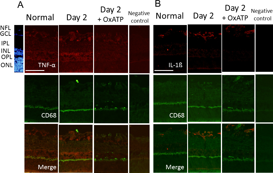
 Figure 5 of
Sugiyama, Mol Vis 2013; 19:2080-2091.
Figure 5 of
Sugiyama, Mol Vis 2013; 19:2080-2091.